73 7.6 Human Body Cavities
Created by CK-12 Foundation

Contain the Brain
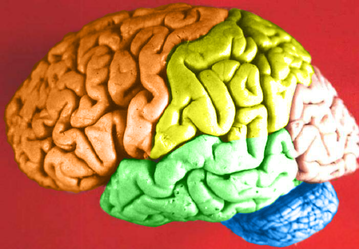
You probably recognize the colourful object in this photo (Figure 7.6.1) as a human brain. The brain is arguably the most important organ in the human body. Fortunately for us, the brain has its own special “container,” called the cranial cavity. The cranial cavity enclosing the brain is just one of several cavities in the human body that form “containers” for vital organs.
What Are Body Cavities?
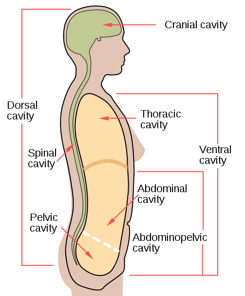
The human body, like that of many other multicellular organisms, is divided into a number of body cavities. A body cavity is a fluid-filled space inside the body that holds and protects internal organs. Human body cavities are separated by membranes and other structures. The two largest human body cavities are the ventral cavity and dorsal cavity. These two body cavities are subdivided into smaller body cavities. Both the dorsal and ventral cavities and their subdivisions are shown in the Figure 7.6.2 diagram.

Ventral Cavity
The ventral cavity is at the anterior (or front) of the trunk. Organs contained within this body cavity include the lungs, heart, stomach, intestines, and reproductive organs. The ventral cavity allows for considerable changes in the size and shape of the organs inside as they perform their functions. Organs such as the lungs, stomach, or uterus, for example, can expand or contract without distorting other tissues or disrupting the activities of nearby organs.
The ventral cavity is subdivided into the thoracic and abdominopelvic cavities.
- The thoracic cavity fills the chest and is subdivided into two pleural cavities and the pericardial cavity. The pleural cavities hold the lungs, and the pericardial cavity holds the heart.
- The abdominopelvic cavity fills the lower half of the trunk and is subdivided into the abdominal cavity and the pelvic cavity. The abdominal cavity holds digestive organs and the kidneys, and the pelvic cavity holds reproductive organs and organs of excretion.
Dorsal Cavity
The dorsal cavity is at the posterior (or back) of the body, including both the head and the back of the trunk. The dorsal cavity is subdivided into the cranial and spinal cavities.
- The cranial cavity fills most of the upper part of the skull and contains the brain.
- The spinal cavity is a very long, narrow cavity inside the vertebral column. It runs the length of the trunk and contains the spinal cord.
The brain and spinal cord are protected by the bones of the skull and the vertebrae of the spine. They are further protected by the meninges, a three-layer membrane that encloses the brain and spinal cord. A thin layer of cerebrospinal fluid is maintained between two of the meningeal layers. This clear fluid is produced by the brain, and it provides extra protection and cushioning for the brain and spinal cord.
Feature: My Human Body
The meninges membranes that protect the brain and spinal cord inside their cavities may become inflamed, generally due to a bacterial or viral infection. This condition is called meningitis, and it can lead to serious long-term consequences such as deafness, epilepsy, or cognitive deficits, especially if not treated quickly. Meningitis can also rapidly become life-threatening, so it is classified as a medical emergency.
Learning the symptoms of meningitis may help you or a loved one get prompt medical attention if you ever develop the disease. Common symptoms include fever, headache, and neck stiffness. Other symptoms include confusion or altered consciousness, vomiting, and an inability to tolerate light or loud noises. Young children often exhibit less specific symptoms, such as irritability, drowsiness, or poor feeding.
Meningitis is diagnosed with a lumbar puncture (commonly known as a “spinal tap”), in which a needle is inserted into the spinal canal to collect a sample of cerebrospinal fluid. The fluid is analyzed in a medical lab for the presence of pathogens. If meningitis is diagnosed, treatment consists of antibiotics and sometimes antiviral drugs. Corticosteroids may also be administered to reduce inflammation and the risk of complications (such as brain damage). Supportive measures such as IV fluids may also be provided.
Some types of meningitis can be prevented with a vaccine. Ask your health care professional whether you have had the vaccine or should get it. Giving antibiotics to people who have had significant exposure to certain types of meningitis may reduce their risk of developing the disease. If someone you know is diagnosed with meningitis and you are concerned about contracting the disease yourself, see your doctor for advice.
7.6 Summary
- The human body is divided into a number of body cavities, fluid-filled spaces in the body that hold and protect internal organs. The two largest human body cavities are the ventral cavity and dorsal cavity.
- The ventral cavity is at the anterior (or front) of the trunk. It is subdivided into the thoracic cavity and abdominopelvic cavity.
- The dorsal cavity is at the posterior (or back) of the body, and includes the head and the back of the trunk. It is subdivided into the cranial cavity and spinal cavity.
7.6 Review Questions
-
- What is a body cavity?
- Compare and contrast the ventral and dorsal body cavities.
- Identify the subdivisions of the ventral cavity, and the organs each contains.
- Describe the subdivisions of the dorsal cavity and their contents.
- Identify and describe all the tissues that protect the brain and spinal cord.
- What do you think might happen if fluid were to build up excessively in one of the body cavities?
- Explain why a woman’s body can accommodate a full-term fetus during pregnancy without damaging her internal organs.
- Which body cavity does the needle enter in a lumbar puncture?
- What are the names given to the three body cavity divisions where the heart is located?What are the names given to the three body cavity divisions where the kidneys are located?
-
7.6 Explore More
Why is meningitis so dangerous? – Melvin Sanicas, TED-Ed, 2018.
Attributions
Figure 7.6.1
Brain Lobes by John A Beal, Department of Cellular Biology & Anatomy, Louisiana State University Health Sciences Center Shreveport on Wikimedia Commons is used under a CC BY 2.5 (https://creativecommons.org/licenses/by/2.5/deed.en) license.
Figure 7.6.2
body_cavities-en.svg by Mysid (SVG) on Wikimedia Commons is in the public domain (https://en.wikipedia.org/wiki/Public_domain). (Original by US National Cancer Institute [NCI].)
References
Mayo Clinic Staff. (n.d.). Meningitis. MayoClinic.org. https://www.mayoclinic.org/diseases-conditions/meningitis/symptoms-causes/syc-20350508
TED-Ed. (2018, November 19). Why is meningitis so dangerous? – Melvin Sanicas. YouTube. https://www.youtube.com/watch?v=IaQdv_dBDqM&feature=youtu.be
A fluid-filled space inside the body that holds and protects internal organs.
A major human body cavity at the anterior (front) of the trunk that contains such organs as the lungs, heart, stomach, intestines, and internal reproductive organs.
A body cavity in the chest that holds the lungs and heart.
Body cavity that fills the lower half of the trunk and holds the kidneys and the digestive and reproductive organs.
A major human body cavity that includes the head and the posterior (back) of the trunk and holds the brain and spinal cord.
A cavity that fills most of the upper part of the skull and contains the brain.
A long, narrow body cavity inside the vertebral column that runs the length of the trunk and contains the spinal cord.
A three-layered membrane that encloses and protects the brain and spinal cord and contains cerebrospinal fluid.
Clear fluid produced by the brain that forms a thin layer within the meninges and provides protection and cushioning for the brain and spinal cord.

