
111 12.3 Types of Muscle Tissue
Created by CK-12 Foundation/Adapted by Christine Miller

Work Those Eye Muscles!
Imagine the man in Figure 12.3.1 turns his eyes in your direction. This is a very small movement, considering the conspicuously large and strong external eye muscles that control eyeball movements. These muscles have been called the strongest muscles in the human body relative to the work they do. However, the external eye muscles actually do a surprising amount of work. Eye movements occur almost constantly during waking hours, especially when we are scanning faces or reading. Eye muscles are also exercised nightly during the phase of sleep called rapid eye movement sleep. External eye muscles can move the eyes because they are made mainly of muscle tissue.
What is Muscle Tissue?
Muscle tissue is a soft tissue that makes up most of the tissues in the muscles of the human muscular system. Other tissues in muscles are connective tissues, such as tendons that attach skeletal muscles to bones and sheaths of connective tissues that cover or line muscle tissues. Only muscle tissue per se, has cells with the ability to contract.
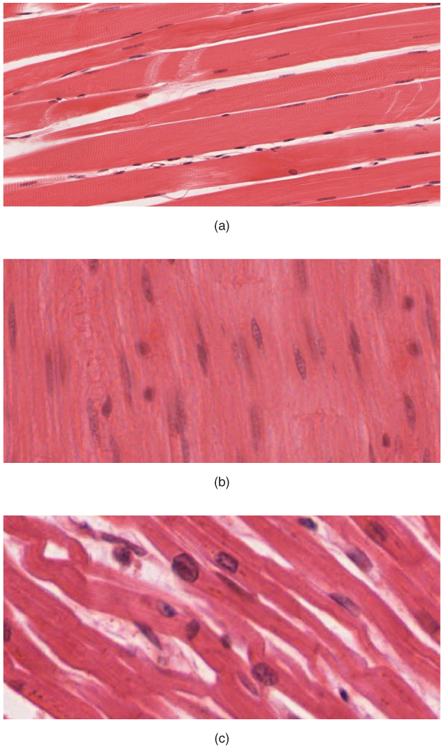
There are three major types of muscle tissues in the human body: skeletal, smooth, and cardiac muscle tissues. Figure 12.3.2 shows how the three types of muscle tissues appear under magnification. When you read about each type below, you will learn why the three types appear as they do.
Skeletal Muscle Tissue
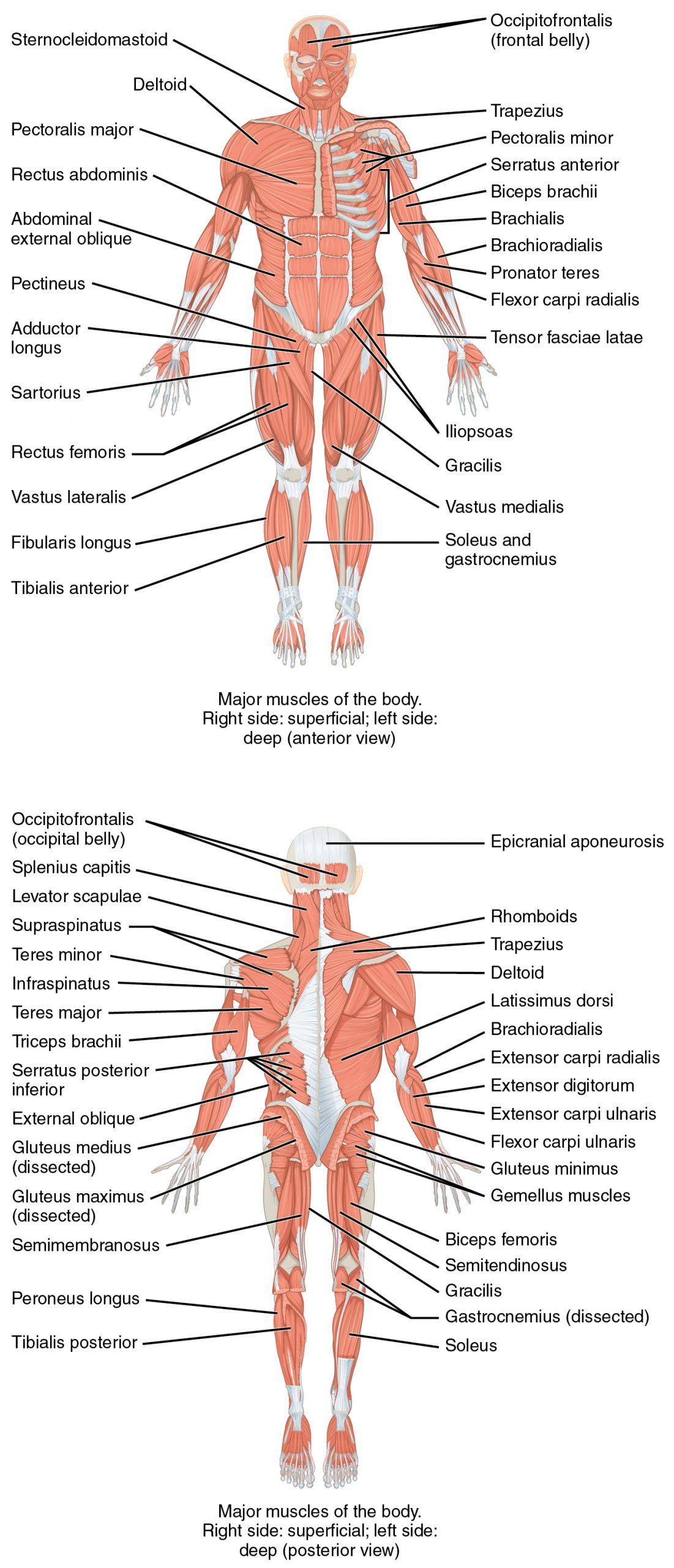
Skeletal muscle is muscle tissue that is attached to bones by tendons, which are bundles of collagen fibres. Whether you are moving your eyes or running a marathon, you are using skeletal muscles. Contractions of skeletal muscles are voluntary, or under conscious control of the central nervous system via the somatic nervous system. Skeletal muscle tissue is the most common type of muscle tissue in the human body. By weight, an average adult male is about 42% skeletal muscles, and the average adult female is about 36% skeletal muscles. Some of the major skeletal muscles in the human body are labeled in Figure 12.3.3 below.
Skeletal Muscle Pairs
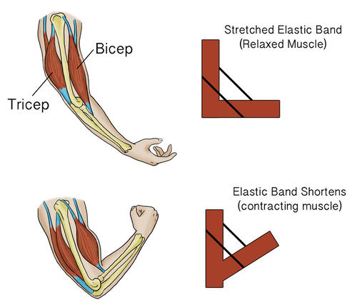
To move bones in opposite directions, skeletal muscles often consist of muscle pairs that work in opposition to one another, also called antagonistic muscle pairs. For example, when the biceps muscle (on the front of the upper arm) contracts, it can cause the elbow joint to flex or bend the arm, as shown in Figure 12.3.4. When the triceps muscle (on the back of the upper arm) contracts, it can cause the elbow to extend or straighten the arm. The biceps and triceps muscles, also shown in Figure 12.3.4, are an example of a muscle pair where the muscles work in opposition to each other.
Skeletal Muscle Structure
Each skeletal muscle consists of hundreds — or even thousands — of skeletal muscle fibres, which are long, string-like cells. As shown in Figure 12.3.5 below, skeletal muscle fibres are individually wrapped in connective tissue called endomysium. The skeletal muscle fibres are bundled together in units called muscle fascicles, which are surrounded by sheaths of connective tissue called perimysium. Each fascicle contains between ten and 100 (or even more!) skeletal muscle fibres. Fascicles, in turn, are bundled together to form individual skeletal muscles, which are wrapped in connective tissue called epimysium. The connective tissues in skeletal muscles have a variety of functions. They support and protect muscle fibres, allowing them to withstand the forces of contraction by distributing the forces applied to the muscle. They also provide pathways for nerves and blood vessels to reach the muscles. In addition, the epimysium anchors the muscles to tendons.
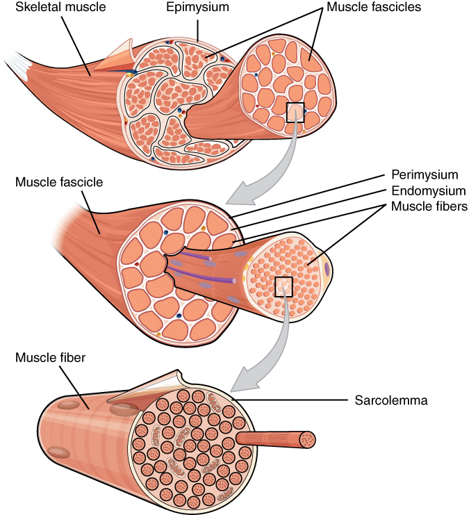
The same bundles-within-bundles structure is replicated within each muscle fibre. As shown in Figure 12.3.6, a muscle fibre consists of a bundle of myofibrils, which are themselves bundles of protein filaments. These protein filaments consist of thin filaments of the protein actin, which are anchored to structures called Z discs, and thick filaments of the protein myosin. The filaments are arranged together within a myofibril in repeating units called sarcomeres, which run from one Z disc to the next. The sarcomere is the basic functional unit of skeletal and cardiac muscles. It contracts as actin and myosin filaments slide over one another. Skeletal muscle tissue is said to be striated, because it appears striped. It has this appearance because of the regular, alternating A (dark) and I (light) bands of filaments arranged in sarcomeres inside the muscle fibres. Other components of a skeletal muscle fibre include multiple nuclei and mitochondria.

Slow- and Fast-Twitch Skeletal Muscle Fibres
Skeletal muscle fibres can be divided into two types, called slow-twitch (or type I) muscle fibres and fast-twitch (or type II) muscle fibres.
- Slow-twitch muscle fibres are dense with capillaries and rich in mitochondria and myoglobin, which is a protein that stores oxygen until needed for muscle activity. Relative to fast-twitch fibres, slow-twitch fibres can carry more oxygen and sustain aerobic (oxygen-using) activity. Slow-twitch fibres can contract for long periods of time, but not with very much force. They are relied upon primarily in endurance events, such as distance running or cycling.
- Fast-twitch muscle fibres contain fewer capillaries and mitochondria and less myoglobin. This type of muscle fibre can contract rapidly and powerfully, but it fatigues very quickly. Fast-twitch fibres can sustain only short, anaerobic (non-oxygen-using) bursts of activity. Relative to slow-twitch fibres, fast-twitch fibres contribute more to muscle strength and have a greater potential for increasing in mass. They are relied upon primarily in short, strenuous events, such as sprinting or weightlifting.
Proportions of fibre types vary considerably from muscle to muscle and from person to person. Individuals may be genetically predisposed to have a larger percentage of one type of muscle fibre than the other. Generally, an individual who has more slow-twitch fibres is better suited for activities requiring endurance, whereas an individual who has more fast-twitch fibres is better suited for activities requiring short bursts of power.
Smooth Muscle
Smooth muscle is muscle tissue in the walls of internal organs and other internal structures such as blood vessels. When smooth muscles contract, they help the organs and vessels carry out their functions. When smooth muscles in the stomach wall contract, for example, they squeeze the food inside the stomach, helping to mix and churn the food and break it into smaller pieces. This is an important part of digestion. Contractions of smooth muscles are involuntary, so they are not under conscious control. Instead, they are controlled by the autonomic nervous system, hormones, neurotransmitters, and other physiological factors.
Structure of Smooth Muscle
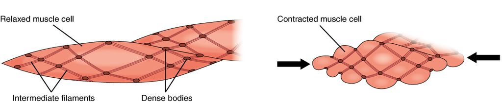
The cells that make up smooth muscle are generally called myocytes. Unlike the muscle fibres of striated muscle tissue, the myocytes of smooth muscle tissue do not have their filaments arranged in sarcomeres. Therefore, smooth tissue is not striated. However, the myocytes of smooth muscle do contain myofibrils, which in turn contain bundles of myosin and actin filaments. The filaments cause contractions when they slide over each other, as shown in Figure 12.3.7.
Functions of Smooth Muscle
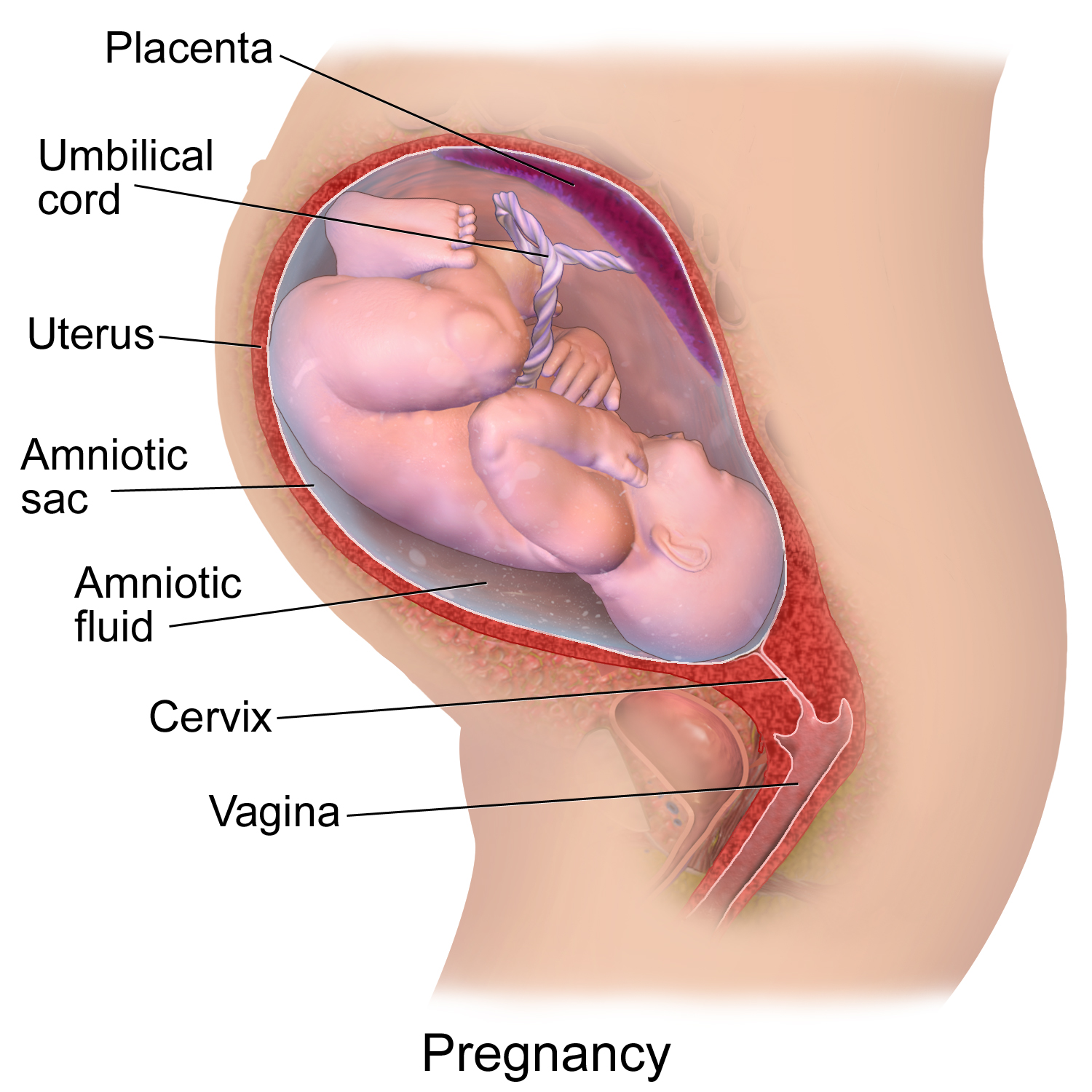
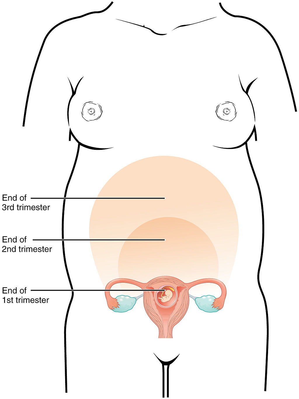
Unlike striated muscle, smooth muscle can sustain very long-term contractions. Smooth muscle can also stretch and still maintain its contractile function, which striated muscle cannot. The elasticity of smooth muscle is enhanced by an extracellular matrix secreted by myocytes. The matrix consists of elastin, collagen, and other stretchy fibres. The ability to stretch and still contract is an important attribute of smooth muscle in organs such as the stomach and uterus (see Figures 12.3.8 and 12.3.9), both of which must stretch considerably as they perform their normal functions.
 |
 |
The following list indicates where many smooth muscles are found, along with some of their specific functions.
- Walls of organs of the gastrointestinal tract (such as the esophagus, stomach, and intestines), moving food through the tract by peristalsis
- Walls of air passages of the respiratory tract (such as the bronchi), controlling the diameter of the passages and the volume of air that can pass through them
- Walls of organs of the male and female reproductive tracts; in the uterus, for example, pushing a baby out of the uterus and into the birth canal
- Walls of structures of the urinary system, including the urinary bladder, allowing the bladder to expand so it can hold more urine, and then contract as urine is released
- Walls of blood vessels, controlling the diameter of the vessels and thereby affecting blood flow and blood pressure
- Walls of lymphatic vessels, squeezing the fluid called lymph through the vessels
- Iris of the eyes, controlling the size of the pupils and thereby the amount of light entering the eyes
- Arrector pili in the skin, raising hairs in hair follicles in the dermis
Cardiac Muscle
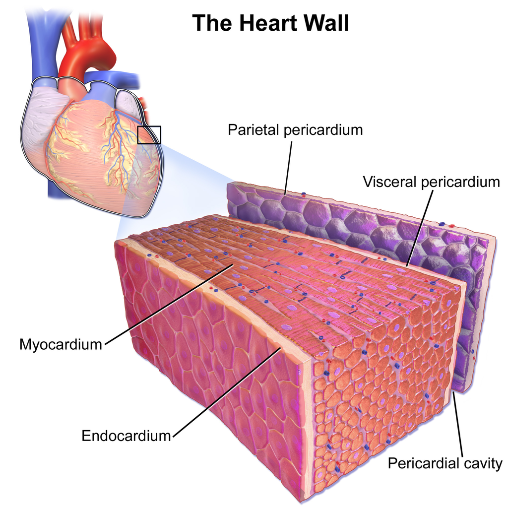
Cardiac muscle is found only in the wall of the heart. It is also called myocardium. As shown in Figure 12.3.10, myocardium is enclosed within connective tissues, including the endocardium on the inside of the heart and pericardium on the outside of the heart. When cardiac muscle contracts, the heart beats and pumps blood. Contractions of cardiac muscle are involuntary, like those of smooth muscles. They are controlled by electrical impulses from specialized cardiac muscle cells in an area of the heart muscle called the sinoatrial node.
Like skeletal muscle, cardiac muscle is striated because its filaments are arranged in sarcomeres inside the muscle fibres. However, in cardiac muscle, the myofibrils are branched at irregular angles rather than arranged in parallel rows (as they are in skeletal muscle). This explains why cardiac and skeletal muscle tissues look different from one another.
The cells of cardiac muscle tissue are arranged in interconnected networks. This arrangement allows rapid transmission of electrical impulses, which stimulate virtually simultaneous contractions of the cells. This enables the cells to coordinate contractions of the heart muscle.
The heart is the muscle that performs the greatest amount of physical work in the course of a lifetime. Although the power output of the heart is much less than the maximum power output of some other muscles in the human body, the heart does its work continuously over an entire lifetime without rest. Cardiac muscle contains a great many mitochondria, which produce ATP for energy and help the heart resist fatigue.
Feature: Human Biology in the News
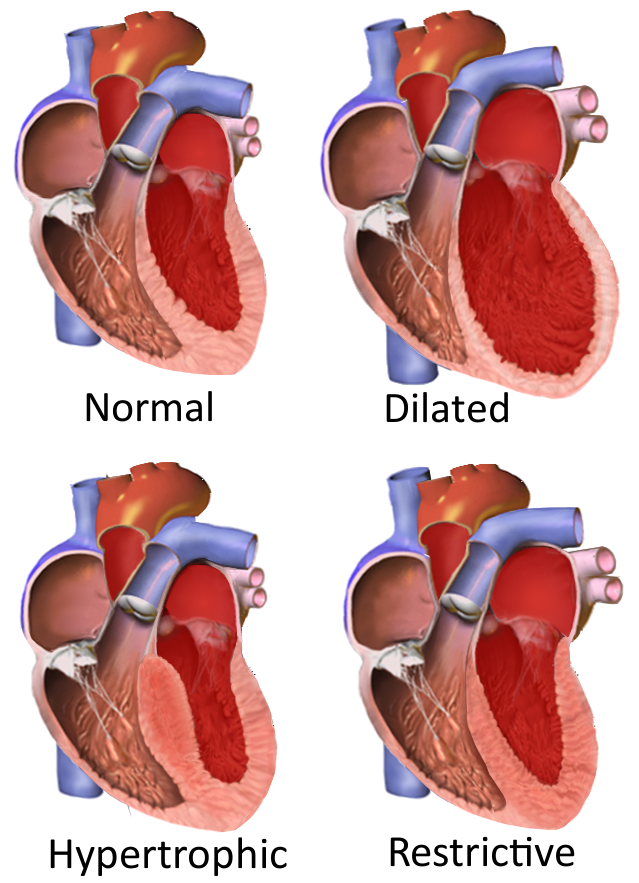
Cardiomyopathy is a disease in which the muscles of the heart are no longer able to effectively pump blood to the body — extreme forms of this disease can lead to heart failure. There are four main types of cardiomyopathy (also illustrated in Figure 12.3.11):
- Dilated (congestive) cardiomyopathy: the left ventricle (the chamber itself) of the heart becomes enlarged and can’t pump blood our to the body. This is normally related to coronary artery disease and/or heart attack
- Hypertrophic cardiomyopathy: abnormal thickening of the muscular walls of the left ventricle make the chamber less able to work properly. This condition is more common in patients with a family history of the disease.
- Restrictive cardiomyopathy: the myocardium becomes abnormally rigid and inelastic and is unable to expand in between heartbeats to refill with blood. Restrictive cardiomyopathy typically affects older people.
- Arrhythmogenic right ventricular cardiomyopathy: the right ventricular muscle is replaced by adipose or scar tissue, reducing elasticity and interfering with normal heartbeat and rhythm. This disease is often caused by genetic mutations.
Cardiomyopathy is typically diagnosed with a physical exam supplemented by medical and family history, an angiogram, blood tests, chest x-rays and electrocardiograms. In some cases your doctor would also requisition a CT scan and/or genetic testing.
When treating cardiomyopathy, the goal is to reduce symptoms that affect everyday life. Certain medications can help regularize and slow heart rate, decrease chances of blood clots and cause vasodilation in the coronary arteries. If medication is not sufficient to manage symptoms, a pacemaker or even a heart transplant may be the best option. Lifestyle can also help manage the symptoms of cardiomyopathy — people living with this disease are encouraged to avoid drug and alcohol use, control high blood pressure, eat a healthy diet, get ample rest and exercise, as well as reduce stress levels.
12.3 Summary
- Muscle tissue is a soft tissue that makes up most of the tissues in the muscles of the human muscular system. It is the only type of tissue that has cells with the ability to contract.
- Skeletal muscle tissue is attached to bones by tendons. It allows voluntary body movements.
- Skeletal muscle is the most common type of muscle tissue in the human body. To move bones in opposite directions, skeletal muscles often consist of pairs of muscles that work in opposition to one another to move bones in different directions at joints.
- Skeletal muscle fibres are bundled together in units called muscle fascicles, which are bundled together to form individual skeletal muscles. Skeletal muscles also have connective tissue supporting and protecting the muscle tissue.
- Each skeletal muscle fibre consists of a bundle of myofibrils, which are bundles of protein filaments. The filaments are arranged in repeating units called sarcomeres, which are the basic functional units of skeletal muscles. Skeletal muscle tissue is striated because of the pattern of sarcomeres in its fibres.
- Skeletal muscle fibres can be divided into two types, called slow-twitch and fast-twitch fibres. Slow-twitch fibres are used mainly in aerobic endurance activities, such as long-distance running. Fast-twitch fibres are used mainly for non-aerobic, strenuous activities, such as sprinting. Proportions of the two types of fibres vary from muscle to muscle and person to person.
- Smooth muscle tissue is found in the walls of internal organs and vessels. When smooth muscles contract, they help the organs and vessels carry out their functions. Contractions of smooth muscles are involuntary and controlled by the autonomic nervous system, hormones, and other substances.
- Cells of smooth muscle tissue are not striated because they lack sarcomeres, but the cells contract in the same basic way as striated muscle cells. Unlike striated muscle, smooth muscle can sustain very long-term contractions and maintain its contractile function, even when stretched.
- Cardiac muscle tissue is found only in the wall of the heart. When cardiac muscle contracts, the heart beats and pumps blood. Contractions of cardiac muscle are involuntary, like those of smooth muscles. They are controlled by electrical impulses from specialized cardiac cells.
- Like skeletal muscle, cardiac muscle is striated because its filaments are arranged in sarcomeres inside the muscle fibres. However, the myofibrils are branched instead of arranged in parallel rows, making cardiac and skeletal muscle tissues look different from one another.
- The heart is the muscle that performs the greatest amount of physical work in the course of a lifetime. Its cells contain a great many mitochondria to produce ATP for energy and help the heart resist fatigue.
12.3 Review Questions
- What is muscle tissue?
-
-
- Where is skeletal muscle found, and what is its general function?
- Why do many skeletal muscles work in pairs?
- Describe the structure of a skeletal muscle.
- Relate muscle fibre structure to the functional units of muscles.
- Why is skeletal muscle tissue striated?
- Where is smooth muscle found? What controls the contraction of smooth muscle?
- Where is cardiac muscle found? What controls its contractions?
- The heart muscle is smaller and less powerful than some other muscles in the body. Why is the heart the muscle that performs the greatest amount of physical work in the course of a lifetime? How does the heart resist fatigue?
- Give one example of connective tissue that is found in muscles. Describe one of its functions.
12.3 Explore More
What happens during a heart attack? – Krishna Sudhir, TED-Ed, 2017.
Three types of muscle | Circulatory system physiology | NCLEX-RN | KhanAcademyMedicine, 2012.
Attributions
Figure 12.3.1
Look by ali-yahya-155huuQwGvA [photo] by Ali Yahya on Unsplash is used under the Unsplash License (https://unsplash.com/license).
Figure 12.3.2
Skeletal_Smooth_Cardiac by OpenStax College on Wikimedia Commons is used under a CC BY 3.0 (https://creativecommons.org/licenses/by/3.0) license.
Figure 12.3.3
Anterior_and_Posterior_Views_of_Muscles by OpenStax on Wikimedia Commons is used under a CC BY 4.0 (https://creativecommons.org/licenses/by/4.0) license.
Figure 12.3.4
Antagonistic Muscle Pair by Laura Guerin at CK-12 Foundation on Wikimedia Commons is used under a CC BY-NC 3.0 (https://creativecommons.org/licenses/by-nc/3.0/) license.
Figure 12.3.5
Muscle_Fibes_(large) by OpenStax on Wikimedia Commons is used under a CC BY 4.0 (https://creativecommons.org/licenses/by/4.0) license.
Figure 12.3.6
Muscle_Fibers_(small) by OpenStax on Wikimedia Commons is used under a CC BY 4.0 (https://creativecommons.org/licenses/by/4.0) license.
Figure 12.3.7
Smooth_Muscle_Contraction by OpenStax on Wikimedia Commons is used under a CC BY 4.0 (https://creativecommons.org/licenses/by/4.0) license.
Figure 12.3.8
Blausen_0747_Pregnancy by BruceBlaus on Wikimedia Commons is used under a CC BY 3.0 (https://creativecommons.org/licenses/by/3.0) license.
Figure 12.3.9
Size_of_Uterus_Throughout_Pregnancy-02 by OpenStax College on Wikimedia Commons is used under a CC BY 3.0 (https://creativecommons.org/licenses/by/3.0) license.
Figure 12.3.10
1024px-Blausen_0470_HeartWall by BruceBlaus on Wikimedia Commons is used under a CC BY 3.0 (https://creativecommons.org/licenses/by/3.0) license.
Figure 12.3.11
Tipet_e_kardiomiopative by Npatchett at English Wikipedia on Wikimedia Commons is used under a CC BY-SA 3.0 (https://creativecommons.org/licenses/by-sa/3.0) license. (Work derived from Blausen 0165 Cardiomyopathy Dilated by BruceBlaus)
References
Betts, J. G., Young, K.A., Wise, J.A., Johnson, E., Poe, B., Kruse, D.H., Korol, O., Johnson, J.E., Womble, M., DeSaix, P. (2013, June 19). Figure 4.18 Muscle tissue [digital image]. In Anatomy and Physiology (Section 4.4). OpenStax. https://openstax.org/books/anatomy-and-physiology/pages/4-4-muscle-tissue-and-motion
Betts, J. G., Young, K.A., Wise, J.A., Johnson, E., Poe, B., Kruse, D.H., Korol, O., Johnson, J.E., Womble, M., DeSaix, P. (2013, June 19). Figure 28.18 Size of uterus throughout pregnancy [digital image]. In Anatomy and Physiology (Section 28.4). OpenStax. https://openstax.org/books/anatomy-and-physiology/pages/28-4-maternal-changes-during-pregnancy-labor-and-birth
Betts, J. G., Young, K.A., Wise, J.A., Johnson, E., Poe, B., Kruse, D.H., Korol, O., Johnson, J.E., Womble, M., DeSaix, P. (2016, May 18). Figure 10.3 The three connective tissue layers [digital image]. In Anatomy and Physiology (Section 10.2). OpenStax. https://openstax.org/books/anatomy-and-physiology/pages/10-2-skeletal-muscle
Betts, J. G., Young, K.A., Wise, J.A., Johnson, E., Poe, B., Kruse, D.H., Korol, O., Johnson, J.E., Womble, M., DeSaix, P. (2016, May 18). Figure 10.4 Muscle fiber [digital image]. In Anatomy and Physiology (Section 10.2). OpenStax. https://openstax.org/books/anatomy-and-physiology/pages/10-2-skeletal-muscle
Betts, J. G., Young, K.A., Wise, J.A., Johnson, E., Poe, B., Kruse, D.H., Korol, O., Johnson, J.E., Womble, M., DeSaix, P. (2016, May 18). Figure 10.24 Muscle contraction [digital image]. In Anatomy and Physiology (Section 10.8). OpenStax. https://openstax.org/books/anatomy-and-physiology/pages/10-8-smooth-muscle
Betts, J. G., Young, K.A., Wise, J.A., Johnson, E., Poe, B., Kruse, D.H., Korol, O., Johnson, J.E., Womble, M., DeSaix, P. (2016, May 18). Figure 11.5 Overview of the muscular system [digital image]. In Anatomy and Physiology (Section 11.2). OpenStax. https://openstax.org/books/anatomy-and-physiology/pages/11-2-naming-skeletal-muscles
Blausen.com staff. (2014). Medical gallery of Blausen Medical 2014. WikiJournal of Medicine 1 (2). DOI:10.15347/wjm/2014.010. ISSN 2002-4436.
Brainard, J/ CK-12 Foundation. (2012). Figure 5 Triceps and biceps muscles in the upper arm are opposing muscles. [digital image]. In CK-12 Biology (Section 21.3) [online Flexbook]. CK12.org. https://www.ck12.org/book/ck-12-biology/section/21.3/ (Last modified August 11, 2017.)
khanacademymedicine. (2012, October 19). Three types of muscle | Circulatory system physiology | NCLEX-RN | Khan Academy. YouTube.
TED-Ed. (2017, February 14). What happens during a heart attack? – Krishna Sudhir. YouTube. https://www.youtube.com/watch?v=3_PYnWVoUzM&feature=youtu.be
A soft tissue that composes muscles in animal bodies, and gives rise to muscles' ability to contract. This is opposed to other components or tissues in muscle such as tendons or perimysium.
Dense fibrous connective tissue that attaches skeletal muscle to bones.
Voluntary, striated muscle that is attached to bones of the skeleton and helps the body move.
A rigid organ that constitutes part of the vertebrate skeleton in animals.
One of the four basic types of tissue, connective tissue is found in between other tissues everywhere in the body, including the nervous system and generally forms a framework and support structure for body tissues and organs.
The main structural protein in the extracellular matrix in the various connective tissues in the body. As the main component of connective tissue, it is the most abundant protein in mammals, making up from 25% to 35% of the whole-body protein content.
Actions which take place according to the one's desire or are under control.
One of two main divisions of the nervous system that includes the brain and spinal cord.
A division of the peripheral nervous system that controls voluntary activities.
Meaning within the muscle, is a wispy layer of areolar connective tissue that envelopes each individual muscle fiber. It also contains capillaries, nerves, and lymphatics. It overlies the muscle fiber's cell membrane.
A bundle of skeletal muscle fibers surrounded by perimysium, a type of connective tissue.
The sheath of connective tissue surrounding a bundle of muscle fibers.
A sheath of fibrous elastic tissue surrounding a muscle.
Long filaments that run parallel to each other to form muscle (myo) fibers. The muscle fibers are single multinucleated cells that combine to form the muscle. Myofibrils are made up of repeating subunits called sarcomeres.
A protein that forms (together with myosin) the contractile filaments of muscle cells, and is also involved in motion in other types of cells.
A fibrous protein that forms (together with actin) the contractile filaments of muscle cells and is also involved in motion in other types of cells.
The basic functional unit of skeletal and cardiac muscles, containing actin and myosin protein filaments that slide over one another to produce a shortening of the sarcomere resulting in a muscle contraction.
A type of skeletal muscle cell that is mainly responsible for aerobic activities such as long-distance running.
A double-membrane-bound organelle found in most eukaryotic organisms. Mitochondria convert oxygen and nutrients into adenosine triphosphate (ATP). ATP is the chemical energy "currency" of the cell that powers the cell's metabolic activities.
A type of skeletal muscle cell that is mainly responsible for anaerobic activities such as weight lifting.
An involuntary, nonstriated muscle that is found in the walls of internal organs such as the stomach.
Actions which are not under one's conscious control.
A division of the peripheral nervous system that controls involuntary activities.
A hormone is a signaling molecule produced by glands in multicellular organisms that target distant organs to regulate physiology and behavior.
A type of chemical that transmits signals from the axon of a neuron to another cell across a synapse.
A type of muscle cell that makes up smooth muscle tissue.
A key protein of the extracellular matrix. It is highly elastic and present in connective tissue allowing many tissues in the body to resume their shape after stretching or contracting. Elastin helps skin to return to its original position when it is poked or pinched.
A distinctive pattern of smooth muscle contractions that propels foodstuffs distally through the esophagus and intestines.
Involuntary, striated muscle found only in the walls of the heart; also called myocardium.
The muscular tissue of the heart.
The thin, smooth membrane which lines the inside of the chambers of the heart and forms the surface of the valves.
The membrane enclosing the heart, consisting of an outer fibrous layer and an inner double layer of serous membrane.
A small body of specialized muscle tissue in the wall of the right atrium of the heart that acts as a pacemaker by producing a contractile signal at regular intervals.
A complex organic chemical that provides energy to drive many processes in living cells, e.g. muscle contraction, nerve impulse propagation, and chemical synthesis. Found in all forms of life, ATP is often referred to as the "molecular unit of currency" of intracellular energy transfer.
A structure where two or more bones of the skeleton come together.

.jpg)
.jpg)