
134 15.5 Lower Gastrointestinal Tract
Created by CK-12 Foundation/Adapted by Christine Miller
What Is It?
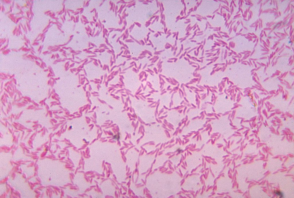
Figure 15.5.1 shows some of the cells of what has been called “the last human organ to be discovered.” This “organ” weighs about 200 grams (about 7 oz) and consists of a hundred trillion cells, yet scientists are only now beginning to learn everything it does and how it varies among individuals. What is it? It’s the mass of bacteria that live in our lower gastrointestinal tract.
Organs of the Lower Gastrointestinal Tract
Most of the bacteria that normally live in the lower gastrointestinal (GI) tract live in the large intestine. They have important and mutually beneficial relationships with the human organism. We provide them with a great place to live, and they provide us with many benefits, some of which you can read about below. Besides the large intestine and its complement of helpful bacteria, the lower GI tract also includes the small intestine. The latter is arguably the most important organ of the digestive system. It is where most chemical digestion and virtually all absorption of nutrients take place.
Small Intestine
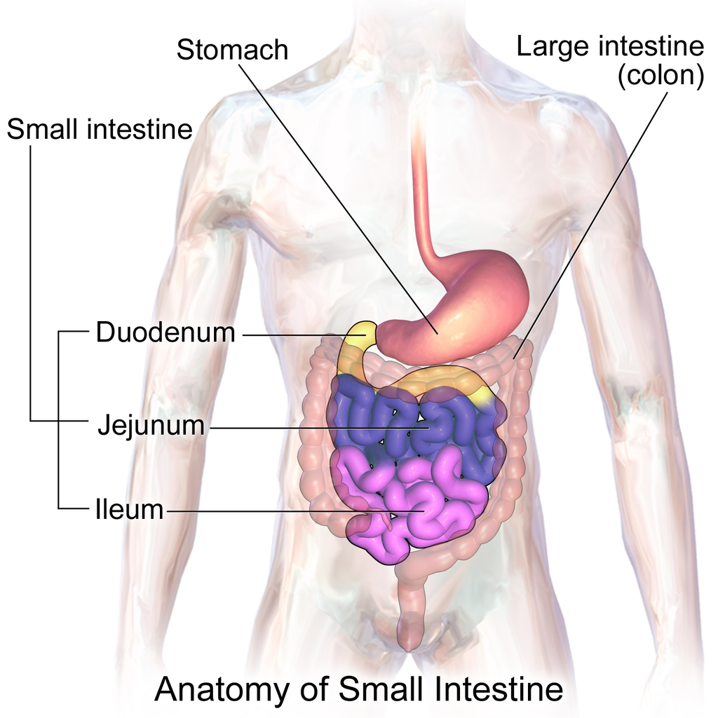
The small intestine (also called the small bowel or gut) is the part of the GI tract between the stomach and large intestine. Its average length in adults is 4.6 m in females and 6.9 m in males. It is approximately 2.5 to 3.0 cm in diameter. It is called “small” because it is much smaller in diameter than the large intestine. The internal surface area of the small intestine totals an average of about 30 m2. Structurally and functionally, the small intestine can be divided into three parts, called the duodenum, jejunum, and ileum, as shown in Figure 15.5.2 and described below.
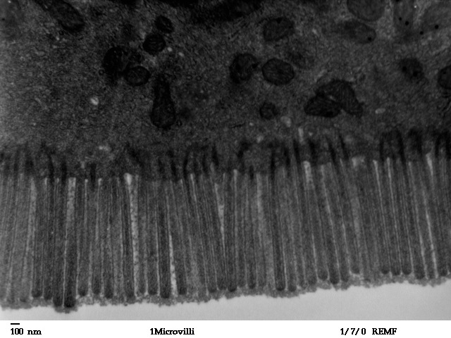
The mucosa lining the small intestine is highly enfolded and covered with the finger-like projections called villi. In fact, each square inch (about 6.5 cm2) of mucosa contains around 20 thousand villi. The individual cells on the surface of the villi also have many finger-like projections — the microvilli, shown in Figure 15.5.3. There are thought to be well over 17 billion microvilli per square centimetre of intestinal mucosa! All of these folds, villi, and microvilli greatly increase the surface area for chyme to come into contact with digestive enzymes, which coat the microvilli, as well as forming a tremendous surface area for the absorption of nutrients. Inside each of the villi is a network of tiny blood and lymph vessels that receive the absorbed nutrients and carry them away in the blood or lymph circulation. The wrinkles and projections in the intestinal mucosa also slow down the passage of chyme so there is more time for digestion and absorption to take place.
Duodenum
The duodenum is the first part of the small intestine, directly connected to the stomach. It is also the shortest part of the small intestine, averaging only about 25 cm (almost 10 in) in length in adults. Its main function is chemical digestion, and it is where most of the chemical digestion in the entire GI tract takes place.
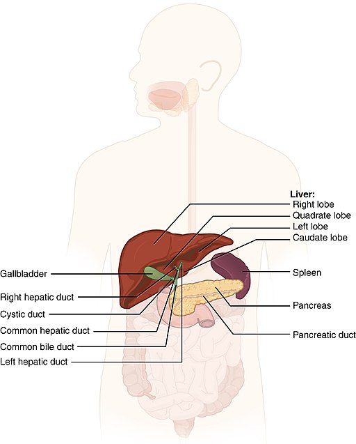
The duodenum receives partially digested, semi-liquid chyme from the stomach. It receives digestive enzymes and alkaline bicarbonate from the pancreas through the pancreatic duct, and it receives bile from the liver via the gallbladder through the common bile duct (see Figure 15.5.4). In addition, the lining of the duodenum secretes digestive enzymes and contains glands — called Brunner’s glands — that secrete mucous and bicarbonate. The bicarbonate from the pancreas and Brunner’s glands — along with bile from the liver — neutralize the highly acidic chyme after it enters the duodenum from the stomach. This is necessary because the digestive enzymes in the duodenum require a nearly neutral environment in order to work. The three major classes of compounds that undergo chemical digestion in the duodenum are carbohydrates, proteins, and lipids.
Digestion of Carbohydrates in the Duodenum
Complex carbohydrates (such as starches) are broken down by the digestive enzyme amylase from the pancreas to short-chain molecules consisting of just a few saccharides (that is, simple sugars). Disaccharides, including sucrose and lactose, are broken down to simple sugars by duodenal enzymes. Sucrase breaks down sucrose, and lactase (if present) breaks down lactose. Some carbohydrates are not digested in the duodenum, and they ultimately pass undigested to the large intestine, where they may be digested by intestinal bacteria.
Digestion of Proteins in the Duodenum
In the duodenum, the pancreatic enzymes trypsin and chymotrypsin cleave proteins into peptides. Then, these smaller molecules are broken down into amino acids. Their digestion is catalyzed by pancreatic enzymes called peptidases.
Digestion of Lipids in the Duodenum
Pancreatic lipase breaks down triglycerides into fatty acids and glycerol. Lipase works with the help of bile secreted by the liver and stored in the gallbladder. Bile salts attach to triglycerides to help them emulsify, or form smaller particles (called micelles) that can disperse through the watery contents of the duodenum. This increases access to the molecules by pancreatic lipase.
Jejunum
The jejunum is the middle part of the small intestine, connecting the duodenum and the ileum. The jejunum is about 2.5 m (about 8 ft) long. Its main function is absorption of the products of digestion, including sugars, amino acids, and fatty acids. Absorption occurs by simple diffusion (water and fatty acids), facilitated diffusion (the simple sugar fructose), or active transport (amino acids, small peptides, water-soluble vitamins, and most glucose). All nutrients are absorbed into the blood, except for fatty acids and fat-soluble vitamins, which are absorbed into the lymph. Although most nutrients are absorbed in the jejunum, there are a few exceptions:
- Iron is absorbed in the duodenum.
- Vitamin B12 and bile salts are absorbed in the ileum.
- Water and lipids are absorbed throughout the small intestine, including the duodenum and ileum in addition to the jejunum.
Ileum
The ileum is the third and final part of the small intestine, directly connected at its distal end to the large intestine. The ileum is about 3 m (almost 10 ft) long. Some cells in the lining of the ileum secrete enzymes that catalyze the final stages of digestion of any undigested protein and carbohydrate molecules. However, the main function of the ileum is to absorb vitamin B12 and bile salts. It also absorbs any other remaining nutrients that were not absorbed in the jejunum. All substances in chyme that remain undigested or unabsorbed by the time they reach the distal end of the ileum pass into the large intestine.
Large Intestine
The large intestine — also called the large bowel — is the last organ of the GI tract. In adults, it averages about 1.5 m (about 5ft) in length. It is shorter than the small intestine, but at least twice as wide, averaging about 6.5 cm (about 2.5 in) in diameter. Water is absorbed from chyme as it passes through the large intestine, turning the chyme into solid feces. Feces is stored in the large intestine until it leaves the body during defecation.
Parts of the Large Intestine
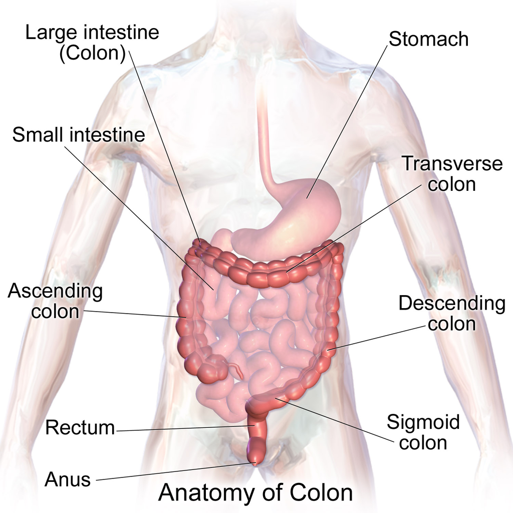
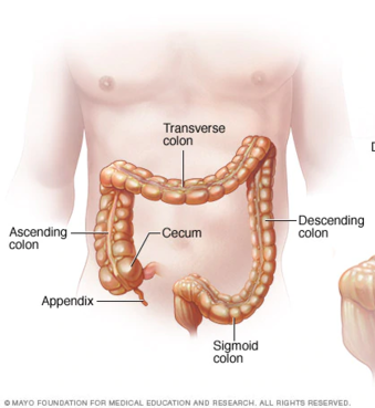
Like the small intestine, the large intestine can be divided into several parts, as shown in Figure 15.5.5. The large intestine begins at the end of the small intestine, where a valve separates the small and large intestines and regulates the movement of chyme into the large intestine. Most of the large intestine is called the colon. The first part of the colon, where chyme enters from the small intestine, is called the cecum. From the cecum, the colon continues upward as the ascending colon, travels across the upper abdomen as the transverse colon, and then continues downward as the descending colon. It then becomes a V-shaped region called the sigmoid colon, which is attached to the rectum. The rectum stores feces until elimination occurs. It transitions to the final part of the large intestine, called the anus, which has an opening to the outside of the body for feces to pass through.
The diagram below (Figure 15.5.6) shows a projection from the cecum of the colon that is known as the appendix. The function of the appendix is somewhat uncertain, but it does not seem to be involved in digestion or absorption. It may play a role in immunity, and in the fetus, it seems to have an endocrine function, releasing hormones needed for homeostasis. Some biologists speculate that the appendix may also store a sample of the colon’s normal bacteria. If so, it may be able to repopulate the colon with the bacteria if illness or antibiotic medications deplete these microorganisms. Appendicitis, or infection and inflammation of the appendix, is a fairly common medical problem, typically resolved by surgical removal of the appendix (appendectomy). People who have had their appendix surgically removed do not seem to suffer any ill effects, so the organ is considered dispensable.
Functions of the Large Intestine
The removal of water from chyme to form feces starts in the ascending colon and continues throughout much of the length of the organ. Salts (such as sodium) are also removed from food wastes in the large intestine before the wastes are eliminated from the body. This allows salts — as well as water — to be recycled in the body.
The large intestine is also the site where huge numbers of beneficial bacteria ferment many unabsorbed materials in food waste. The bacterial breakdown of undigested polysaccharides produces nitrogen, carbon dioxide, methane, and other gases responsible for intestinal gas, or flatulence. These bacteria are particularly prevalent in the descending colon. Some of the bacteria also produce vitamins that are absorbed from the colon. The vitamins include vitamins B1 (thiamine), B2 (riboflavin), B7 (biotin), B12, and K. Another role of bacteria in the colon is an immune function. The bacteria may stimulate the immune system to produce antibodies that are effective against similar, but pathogenic, bacteria, thereby preventing infections. Still other roles played by bacteria in the large intestine include breaking down toxins before they can poison the body, producing substances that help prevent colon cancer, and inhibiting the growth of harmful bacteria.
Feature: My Human Body
Serotonin is a neurotransmitter with a wide variety of functions in the body. Sometimes called the “happy chemical,” it is used in the central nervous system to stabilize mood by contributing to a feeling of well being and happiness. While this “happy chemical” function is important, serotonin is also important for critical brain functions, including support of learning, memory, and reward structures and regulating sleep. Since serotonin’s main target is brain function, you may be surprised to learn that 90% of the human body’s supply of this neurotransmitter is located in the gastrointestinal tract, where it regulates intestinal movements.
The enteric nervous system (ENS), a division of the autonomic nervous system, is a mesh-like system of neurons that control GI function. Interestingly, the ENS can act independently from both the sympathetic/parasympathetic nervous systems and the brain/spinal cord, which is why some scientists refer to the ENS as the “second brain”. The ENS consists of over 500 million neurons (five times as many as in the spinal cord!) and lines the GI tract from the esophagus all the way to the anus. Serotonin is made in and secreted by the gut (small and large intestine) by enterochromaffin cells (also known as Kulchitsky cells) residing in the inner lining of the lower GI tract. While the main function of serotonin in the gut is to regulate digestion, it is also suspected to play a role in brain function — meaning that your gut may actually be partly dictating your mood! Another piece of evidence for the gut-dictating-your-mood theory is the nature vagus nerve, a fibrous visceral nerve, in which 90% of the fibres are dedicated to sending information too the brain, and only 10% of the fibres receiving information from the brain. Some treatments for depression actually involved electrical stimulation of the vagus nerve!
In a 2015 study by researchers at Caltech, it was shown that gut bacteria promote serotonin production by enterochromaffin cells. The study involved measuring the levels of serotonin levels in mice with normal gut bacteria, and then comparing that to the levels of serotonin in a population of mice with no gut bacteria. The germ-free mice were found to produce only 40% of the serotonin that the mice with normal gut flora were producing. Once the germ-free mice were re-colonized with normal gut flora, their production of serotonin returned to normal. This lead researchers to the conclusion that enterochromaffin cells depend on an interaction with gut bacterial flora to be able to produce necessary levels of serotonin.
15.5 Summary
- The lower GI tract includes the small intestine and large intestine. The small intestine is where most chemical digestion and virtually all absorption of nutrients occur. The large intestine contains huge numbers of beneficial bacteria and removes water and salts from food waste before it is eliminated.
- The small intestine consists of three parts: the duodenum, jejunum, and ileum. All three parts of the small intestine are lined with mucosa that is highly enfolded and covered with villi and microvilli, giving the small intestine a huge surface area for digestion and absorption.
- The duodenum secretes digestive enzymes, and also receives bile from the liver via the gallbladder and digestive enzymes and bicarbonate from the pancreas. These digestive substances neutralize acidic chyme and allow for the chemical digestion of carbohydrates, proteins, lipids, and nucleic acids in the duodenum.
- The jejunum carries out most of the absorption of nutrients in the small intestine, including the absorption of simple sugars, amino acids, fatty acids, and many vitamins.
- The ileum carries out any remaining digestion and absorption of nutrients, but its main function is to absorb vitamin B12 and bile salts.
- The large intestine consists of the colon (which, in turn, includes the cecum, ascending colon, transverse colon, descending colon, and sigmoid colon), rectum, and anus. The vestigial organ called the appendix is attached to the cecum of the colon.
- The main function of the large intestine is to remove water and salts from chyme for recycling within the body and eliminating the remaining solid feces from the body through the anus. The large intestine is also the site where trillions of bacteria help digest certain compounds, produce vitamins, stimulate the immune system, and break down toxins, among other important functions.
15.5 Review Questions
-
- How is the mucosa of the small intestine specialized for digestion and absorption?
- What digestive substances are secreted into the duodenum? What compounds in food do they help digest?
- What is the main function of the jejunum?
- What roles does the ileum play?
- How do beneficial bacteria in the large intestine help the human organism?
- When diarrhea occurs, feces leaves the body in a more liquid state than normal. What part of the digestive system do you think is involved in diarrhea? Explain your answer
- What causes intestinal gas, or flatulence?
15.5 Explore More
Why do we pass gas? – Purna Kashyap, TED-Ed, 2014.
What causes constipation? – Heba Shaheed, TED-Ed, 2018.
Attributions
Figure 15.5.1
1024px-Bacteroides_biacutis_01 by CDC/Dr. V.R. Dowell, Jr. Public Health Image Library (PHIL) ID#3087) on Wikimedia Commons is in the public domain (https://en.wikipedia.org/wiki/public_domain).
Figure 15.5.2
Blausen_0817_SmallIntestine_Anatomy by BruceBlaus on Wikimedia Commons is used under a CC BY 3.0 (https://creativecommons.org/licenses/by/3.0) license.
Figure 15.5.3
Human_jejunum_microvilli_1_-_TEM by Louisa Howard, Katherine Connollly / E. M. Facility, Dartmouth, on Wikimedia Commons is in the public domain (https://en.wikipedia.org/wiki/public_domain).
Figure 15.5.4
512px-2422_Accessory_Organs by OpenStax College on Wikimedia Commons is used under a CC BY 3.0 (https://creativecommons.org/licenses/by/3.0) license.
Figure 15.5.5
1024px-Blausen_0603_LargeIntestine_Anatomy by BruceBlaus on Wikimedia Commons is used under a CC BY 3.0 (https://creativecommons.org/licenses/by/3.0) license.
Figure 15.5.6
512px-Ds00070_an01934_im00887_divert_s_gif.webp (1) by Lfreeman04 on Wikimedia Commons is used under a CC BY-SA 4.0 (https://creativecommons.org/licenses/by-sa/4.0) license.
References
Betts, J. G., Young, K.A., Wise, J.A., Johnson, E., Poe, B., Kruse, D.H., Korol, O., Johnson, J.E., Womble, M., DeSaix, P. (2013, June 19). Figure 23.24 Accessory organs [digital image]. In Anatomy and Physiology (Section 23.6). OpenStax. https://openstax.org/books/anatomy-and-physiology/pages/23-6-accessory-organs-in-digestion-the-liver-pancreas-and-gallbladder
Blausen.com Staff. (2014). Medical gallery of Blausen Medical 2014. WikiJournal of Medicine 1 (2). DOI:10.15347/wjm/2014.010. ISSN 2002-4436. /
TED-Ed. (2018, May 7). What causes constipation? – Heba Shaheed. YouTube. https://www.youtube.com/watch?v=0IVO50DuMCs&feature=youtu.be
TED-Ed. (2014, September 8). Why do we pass gas? – Purna Kashyap. YouTube. https://www.youtube.com/watch?v=GTvnjaUU6Xk&feature=youtu.be
Yano, J. M., Yu, K., Donaldson, G. P., Shastri, G. G., Ann, P., Ma, L., Nagler, C. R., Ismagilov, R. F., Mazmanian, S. K., & Hsiao, E. Y. (2015). Indigenous bacteria from the gut microbiota regulate host serotonin biosynthesis. Cell, 161(2), 264–276. https://doi.org/10.1016/j.cell.2015.02.047
The part of the GI tract that includes the small and large intestines.
A long, narrow, tube-like organ of the digestive system where most chemical digestion of food and virtually all absorption of nutrients take place.
Chemical breakdown of large, complex food molecules into smaller, simpler nutrient molecules that can be absorbed by blood or lymph. Usually involves a digestive enzyme.
The first and shortest of three parts of the small intestine where most chemical digestion occurs.
One of three sections that make up the small intestine. The jejunum is located between the duodenum and the ileum.The jejunum makes up about two-fifths of the small intestine. The main function of the jejunum is absorption of important nutrients such as sugars, fatty acids, and amino acids.
The third portion of the small intestine, between the jejunum and the cecum.The ileum helps to further digest food coming from the stomach and other parts of the small intestine.
The innermost tunic of the wall. It lines the lumen of the digestive tract. The mucosa consists of epithelium, an underlying loose connective tissue layer called lamina propria, and a thin layer of smooth muscle called the muscularis mucosa.
A microscopic, finger-like projections in a mucous membrane that form a large surface area for absorption.
One of many tiny projections covering each villus in the mucosal lining the small intestine that increases its absorptive surface.
A thick, semi-liquid mixture that food in the gastrointestinal tract becomes by the time it leaves the stomach.
The measure of how much exposed area a solid object has, expressed in square units.
A hollow, tube-like structure through which blood flows in the cardiovascular system; vein, artery, or capillary.
A sac-like organ of the digestive system between the esophagus and small intestine in which both mechanical and chemical digestion take place.
A long, flat gland that sits tucked behind the stomach in the upper abdomen. The pancreas produces enzymes that help digestion and hormones that help regulate the way your body processes sugar (glucose).
The excretory duct of the pancreas, extending through the gland from tail to head, where it empties into the duodenum.
Fluid produced by the liver and stored in the gall bladder that is secreted into the small intestine to help digest lipids and neutralize acid from the stomach.
An organ of digestion and excretion that secretes bile for lipid digestion and breaks down excess amino acids and toxins in the blood.
A sac-like organ that stores bile from the liver and secretes it into the duodenum of the small intestine as needed for digestion.
A tube that carries bile from the liver and the gallbladder through the pancreas and into the duodenum (the upper part of the small intestine). It is formed where the ducts from the liver and gallbladder are joined. It is part of the biliary duct system.
A slimy substance produced by mucous membranes that traps pathogens, particles, and debris.
A biomolecule consisting of carbon (C), hydrogen (H) and oxygen (O) atoms, usually with a hydrogen–oxygen atom ratio of 2:1. Complex carbohydrates are polymers made from monomers of simple carbohydrates, also termed monosaccharides.
A stored form of glucose used by plants.
An enzyme, found chiefly in saliva and pancreatic fluid, that converts starch and glycogen into simple sugars.
The generic name for sweet-tasting, soluble carbohydrates, many of which are used in food. The various types of sugar are derived from different sources. Simple sugars are called monosaccharides and include glucose, fructose, and galactose.
A digestive enzyme that breaks down proteins in the small intestine. It is secreted by the pancreas in an inactive form, trypsinogen.
A digestive enzyme which breaks down proteins in the small intestine. It is secreted by the pancreas and converted into an active form by trypsin.
A class of biological molecule consisting of linked monomers of amino acids and which are the most versatile macromolecules in living systems and serve crucial functions in essentially all biological processes.
Amino acids are organic compounds that combine to form proteins.
A pancreatic enzyme that catalyzes the breakdown of fats to fatty acids and glycerol.
Process in which substances such as nutrients pass into the blood or lymph.
The movement of a substance from an area of high concentration to an area of low concentration.
The passive movement of molecules across the cell membrane with the aid of a membrane protein.
The movement of ions or molecules across a cell membrane into a region of higher concentration, assisted by enzymes and requiring energy.
A body fluid in humans and other animals that delivers necessary substances such as nutrients and oxygen to the cells and transports metabolic waste products away from those same cells. In vertebrates, it is composed of blood cells suspended in blood plasma.
A fluid that leaks out of capillaries into spaces between cells and circulates in the vessels of the lymphatic system.
An organ of the digestive system that removes water and salts from food waste and forms solid feces for elimination.
Solid waste that remains after food is digested and that is eliminated from the body through the anus.
The discharge of feces from the body... commonly called pooping.
The main part of the large intestine between the small intestine and rectum where water and salts are removed from liquid food wastes to form feces.
A pouch connected to the junction of the small and large intestines.
A short part of the large intestine between the colon and anus where feces is stored until it is eliminated through the anus.
The final part of the large intestine with an opening to the outside for feces to pass through.
A tube-shaped sac attached to and opening into the lower end of the large intestine in humans and some other mammals. Some of the functions of the appendix include maintaining gut flora and immune and lymphatic function.
Any member of a large group of unicellular microorganisms which have cell walls but lack organelles and an organized nucleus, including some which can cause disease.
