
140 16.3 Introduction to the Urinary System
Created by CK-12 Foundation/Adapted by Christine Miller
Figure 16.3.1 The surprising uses of pee.
Surprising Uses
What do gun powder, leather, fabric dyes and laundry service have in common? This may be surprising, but they all historically involved urine. One of the main components in gun powder, potassium nitrate, was difficult to come by pre-1900s, so ingenious gun-owners would evaporate urine to concentrate the nitrates it contains. The ammonium in urine was excellent in breaking down tissues, making it a prime candidate for softening leathers and removing stains in laundry. Ammonia in urine also helps dyes penetrate fabrics, so it was used to make colours stay brighter for longer.
What is the Urinary System?
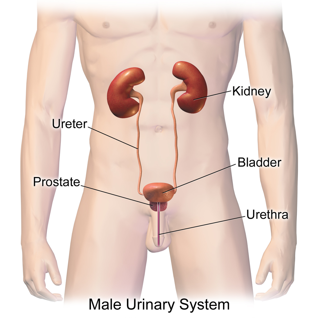
The actual human urinary system, also known as the renal system, is shown in Figure 16.3.2. The system consists of the kidneys, ureters, bladder, and urethra. The main function of the urinary system is to eliminate the waste products of metabolism from the body by forming and excreting urine. Typically, between one and two litres of urine are produced every day in a healthy individual.
Organs of the Urinary System
The urinary system is all about urine. It includes organs that form urine, and also those that transport, store, or excrete urine.
Kidneys
Urine is formed by the kidneys, which filter many substances out of the blood, allow the blood to reabsorb needed materials, and use the remaining materials to form urine. The human body normally has two paired kidneys, although it is possible to get by quite well with just one. As you can see in Figure 16.3.3, each kidney is well supplied with blood vessels by a major artery and vein. Blood to be filtered enters the kidney through the renal artery, and the filtered blood leaves the kidney through the renal vein. The kidney itself is wrapped in a fibrous capsule, and consists of a thin outer layer called the cortex, and a thicker inner layer called the medulla.
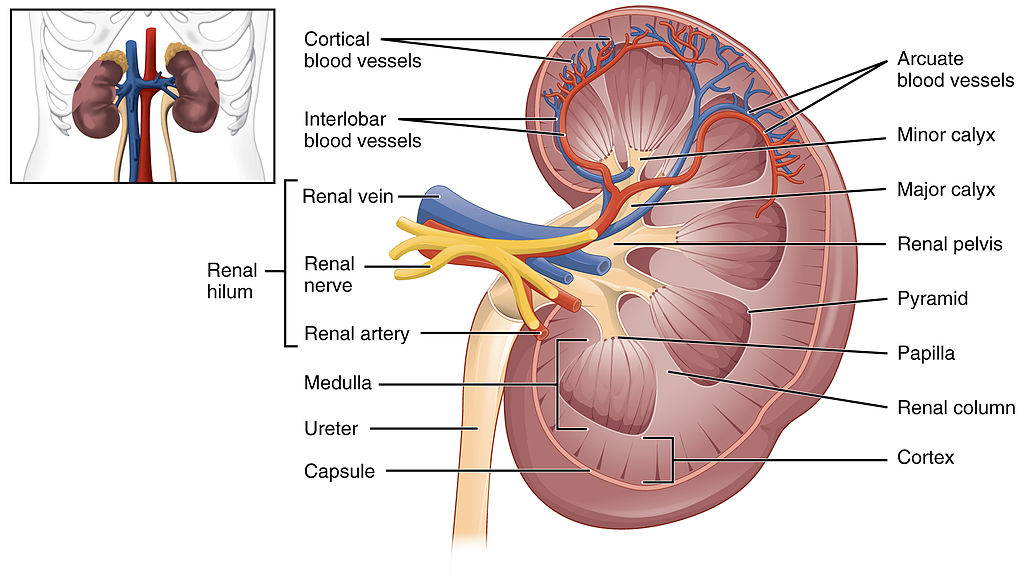
Blood is filtered and urine is formed by tiny filtering units called nephrons. Each kidney contains at least a million nephrons, and each nephron spans the cortex and medulla layers of the kidney. After urine forms in the nephrons, it flows through a system of converging collecting ducts. The collecting ducts join together to form minor calyces (or chambers) that join together to form major calyces (see Figure 16.3.3 above). Ultimately, the major calyces join the renal pelvis, which is the funnel-like end of the ureter where it enters the kidney.
Ureters, Bladder, Urethra
After urine forms in the kidneys, it is transported through the ureters (one per kidney) via peristalsis to the sac-like urinary bladder, which stores the urine until urination. During urination, the urine is released from the bladder and transported by the urethra to be excreted outside the body through the external urethral opening.
Functions of the Urinary System
Waste products removed from the body with the formation and elimination of urine include many water-soluble metabolic products. The main waste products are urea — a by-product of protein catabolism — and uric acid, a by-product of nucleic acid catabolism. Excess water and mineral ions are also eliminated in urine.
Besides the elimination of waste products such as these, the urinary system has several other vital functions. These include:
- Maintaining homeostasis of mineral ions in extracellular fluid: These ions are either excreted in urine or returned to the blood as needed to maintain the proper balance.
- Maintaining homeostasis of blood pH: When pH is too low (blood is too acidic), for example, the kidneys excrete less bicarbonate (which is basic) in urine. When pH is too high (blood is too basic), the opposite occurs, and more bicarbonate is excreted in urine.
- Maintaining homeostasis of extracellular fluids, including the blood volume, which helps maintain blood pressure: The kidneys control fluid volume and blood pressure by excreting more or less salt and water in urine.
Control of the Urinary System
The formation of urine must be closely regulated to maintain body-wide homeostasis. Several endocrine hormones help control this function of the urinary system, including antidiuretic hormone, parathyroid hormone, and aldosterone.
- Antidiuretic hormone (ADH), also called vasopressin, is secreted by the posterior pituitary gland. One of its main roles is conserving body water. It is released when the body is dehydrated, and it causes the kidneys to excrete less water in urine.
- Parathyroid hormone is secreted by the parathyroid glands. It works to regulate the balance of mineral ions in the body via its effects on several organs, including the kidneys. Parathyroid hormone stimulates the kidneys to excrete less calcium and more phosphorus in urine.
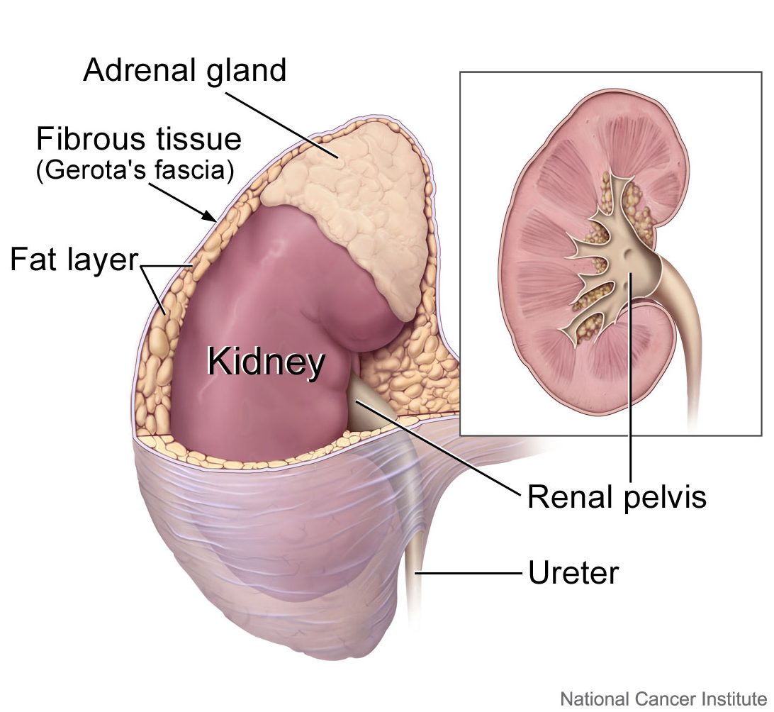
- Aldosterone is secreted by the cortex of the adrenal glands, which rest atop the kidneys, as shown in Figure 16.3.4. Through its effect on the kidneys, it plays a central role in regulating blood pressure. It causes the kidneys to excrete less sodium and water in urine.
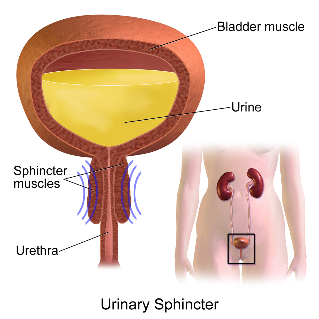
Once urine forms, it is excreted from the body in the process of urination, also sometimes referred to as micturition. This process is controlled by both the autonomic and the somatic nervous systems. As the bladder fills with urine, it causes the autonomic nervous system to signal smooth muscle in the bladder wall to contract (as shown in Figure 16.3.5), and the sphincter between the bladder and urethra to relax and open. This forces urine out of the bladder and through the urethra. Another sphincter at the distal end of the urethra is under voluntary control. When it relaxes under the influence of the somatic nervous system, it allows urine to leave the body through the external urethral opening.
16.3 Summary
- The urinary system consists of the kidneys, ureters, bladder, and urethra. The main function of the urinary system is to eliminate the waste products of metabolism from the body by forming and excreting urine.
- Urine is formed by the kidneys, which filter many substances out of blood, allow the blood to reabsorb needed materials, and use the remaining materials to form urine. Blood to be filtered enters the kidney through the renal artery, and filtered blood leaves the kidney through the renal vein.
- Within each kidney, blood is filtered and urine is formed by tiny filtering units called nephrons, of which there are at least a million in each kidney.
- After urine forms in the kidneys, it is transported through the ureters via peristalsis to the urinary bladder. The bladder stores the urine until urination, when urine is transported by the urethra to be excreted outside the body.
- Besides the elimination of waste products (such as urea, uric acid, excess water, and mineral ions), the urinary system has other vital functions. These include maintaining homeostasis of mineral ions in extracellular fluid, regulating acid-base balance in the blood, regulating the volume of extracellular fluids, and controlling blood pressure.
- The formation of urine must be closely regulated to maintain body-wide homeostasis. Several endocrine hormones help control this function of the urinary system, including antidiuretic hormone from the posterior pituitary gland, parathyroid hormone from the parathyroid glands, and aldosterone from the adrenal glands.
- The process of urination is controlled by both the autonomic and the somatic nervous systems. The autonomic system causes the bladder to empty, but conscious relaxation of the sphincter at the distal end of the urethra allows urine to leave the body.
16.3 Review Questions
-
- State the main function of the urinary system.
- What are nephrons?
- Other than the elimination of waste products, identify functions of the urinary system.
- How is the formation of urine regulated?
- Explain why it is important to have voluntary control over the sphincter at the end of the urethra.
- In terms of how they affect the kidneys, compare aldosterone to antidiuretic hormone.
- If your body needed to retain more calcium, which of the hormones described in this concept is most likely to increase? Explain your reasoning.
16.3 Explore More
The Urinary System – An Introduction | Physiology | Biology | FuseSchool, 2017.
https://youtu.be/pyMcTUQYMQw
Maple Syrup Urine Disease, Alexandria Doody, 2016.
How Accurate Are Drug Tests? Seeker, 2016.
Three Ways Pee Could Change the World, Gross Science, 2015.
Attributions
Figure 16.3.1
- File:Pyrodex powder ffg.jpg by Hustvedt on Wikimedia Commons is used under a CC BY SA 3.0 (https://creativecommons.org/licenses/by-sa/3.0/deed.en).
- Brown leather satchel bag by Álvaro Serrano on Unsplash is used under the Unsplash Licence (https://unsplash.com/license).
- Laundry basket by Andy Fitzsimon on Unsplash is used under the Unsplash Licence (https://unsplash.com/license).
- Tags: Wool Skeins Natural Dyed Colorful Himalayan Weavers by on Pixabay is used under the Pixabay License (https://pixabay.com/service/license/).
Figure 16.3.2
Urinary_System_(Male) by BruceBlaus on Wikimedia Commons is used under a CC BY-SA 4.0 (https://creativecommons.org/licenses/by-sa/4.0) license.
Figure 16.3.3
2610_The_Kidney by OpenStax College on Wikimedia Commons is used under a CC BY 3.0 (https://creativecommons.org/licenses/by/3.0) license.
Figure 16.3.4
Adrenal glands on Kidney by Alan Hoofring (Illustrator)/ NCI Visuals Online is in the public domain (https://en.wikipedia.org/wiki/Public_domain).
Figure 16.3.5
Urinary_Sphincter by BruceBlaus on Wikimedia Commons is used under a CC BY-SA 4.0 (https://creativecommons.org/licenses/by-sa/4.0) license.
References
Alexandria Doody. (2016, March 29). Maple syrup urine disease. YouTube. https://www.youtube.com/watch?v=pyMcTUQYMQw&feature=youtu.be
Betts, J. G., Young, K.A., Wise, J.A., Johnson, E., Poe, B., Kruse, D.H., Korol, O., Johnson, J.E., Womble, M., DeSaix, P. (2013, June 19). Figure 25.8 Left kidney [digital image]. In Anatomy and Physiology (Section 25.3). OpenStax. https://openstax.org/books/anatomy-and-physiology/pages/25-3-gross-anatomy-of-the-kidney
FuseSchool. (2017, June 19). The urinary system – An introduction | Physiology | Biology | FuseSchool. YouTube. https://www.youtube.com/watch?v=dxecGD0m0Xc&feature=youtu.be
Gross Science. (2015, September 15). Three ways pee could change the world. YouTube. https://www.youtube.com/watch?v=xt1Tj5eeS0k&feature=youtu.be
Seeker. (2016, January 16). How accurate are drug tests? YouTube. https://www.youtube.com/watch?v=3z-xjfdJWAI&feature=youtu.be
The body system that produces, stores and eliminates urine, the fluid waste excreted by the kidneys. The kidneys make urine by filtering wastes and extra water from blood. Urine travels from the kidneys through two thin tubes called ureters and fills the bladder.
A liquid waste product of the body that is formed by the kidneys and excreted by the other organs of the urinary system.
One of a pair of organs of the excretory and urinary systems that filters wastes and excess water out of blood and forms urine.
A body fluid in humans and other animals that delivers necessary substances such as nutrients and oxygen to the cells and transports metabolic waste products away from those same cells. In vertebrates, it is composed of blood cells suspended in blood plasma.
One of the million tiny structural and functional units of the kidney that filters blood and forms urine.
The funnel-like end of a ureter where it enters the kidney and where urine collects before it is transported through the ureter.
A muscular, tube-like organ of the urinary system that moves urine by peristalsis from a kidney to the bladder.
A distinctive pattern of smooth muscle contractions that propels foodstuffs distally through the esophagus and intestines.
The process in which urine leaves the body through the external urethral orifice.
Waste product of protein catabolism that is mainly filtered from blood in the kidneys and excreted in urine.
A class of biological molecule consisting of linked monomers of amino acids and which are the most versatile macromolecules in living systems and serve crucial functions in essentially all biological processes.
The breakdown of larger molecules into smaller ones.
A waste product of nucleic acid catabolism that is mainly filtered from blood by the kidneys and excreted in urine.
Large biomolecules, essential to all known forms of life. The term nucleic acid is the overall name for DNA and RNA. They are composed of nucleotides, which are the monomers made of three components: a 5-carbon sugar, a phosphate group and a nitrogenous base.
The body system which acts as a chemical messenger system comprising feedback loops of the hormones released by internal glands of an organism directly into the circulatory system, regulating distant target organs. In humans, the major endocrine glands are the thyroid gland and the adrenal glands.
A hormone is a signaling molecule produced by glands in multicellular organisms that target distant organs to regulate physiology and behavior.
also called vasopressin. A hormone made by the hypothalamus in the brain and stored in the posterior pituitary gland. It tells your kidneys how much water to conserve. ADH constantly regulates and balances the amount of water in your blood.
A hormone secreted by the parathyroid gland which helps regulate blood calcium.
The main mineralocorticoid hormone which is responsible for sodium conservation in the kidney, salivary glands, sweat glands and colon.
division of the peripheral nervous system that controls involuntary activities
A division of the peripheral nervous system that controls voluntary activities.
Actions which take place according to the one's desire or are under control.
The chemical processes that occur in a living organism to sustain life.
A sac-like organ that stores urine until it is excreted from the body.
The ability of an organism to maintain constant internal conditions despite external changes.
The measure of the force exerted by circulating blood on the walls of arteries.
A hormone made by the hypothalamus in the brain and stored in the posterior pituitary gland. It tells your kidneys how much water to conserve. ADH constantly regulates and balances the amount of water in your blood. Higher water concentration increases the volume and pressure of your blood.
The master gland of the endocrine system that secretes many hormones, the majority of which regulate other endocrine glands.
One of a pair of small endocrine glands in the neck that secretes hormones that regulate blood calcium.
one of a pair of glands located on top of the kidneys that secretes hormones such as cortisol and adrenaline
A ring of muscles that can contract to close off an opening between structures, such as between the esophagus and stomach.
A tube-like organ of the urinary system that carries urine out of the body from the bladder and, in males, also carries semen out of the body.

.png)