
126 14.4 Blood Vessels
Created by CK-12 Foundation/Adapted by Christine Miller

Bulging Veins
Why do bodybuilders have such prominent veins? Bulging muscles push surface veins closer to the skin. Combine that with a virtual lack of subcutaneous fat, and you have bulging veins, as well as bulging muscles. Veins are one of three major types of blood vessels in the cardiovascular system.
Types of Blood Vessels
Blood vessels are the part of the cardiovascular system that transports blood throughout the human body. There are three major types of blood vessels. Besides veins, they include arteries and capillaries.
Arteries
Arteries are defined as blood vessels that carry blood away from the heart. Blood flows through arteries largely because it is under pressure from the pumping action of the heart. It should be noted that coronary arteries, which supply heart muscle cells with blood, travel toward the heart, but not as part of the blood flow that travels through the chambers of the heart. Most arteries, including coronary arteries, carry oxygenated blood, but there are a few exceptions, most notably the pulmonary artery. This artery carries deoxygenated blood from the heart to the lungs, where it picks up oxygen and releases carbon dioxide. In virtually all other arteries, the hemoglobin in red blood cells is highly saturated with oxygen (95–100 per cent). These arteries distribute oxygenated blood to tissues throughout the body.
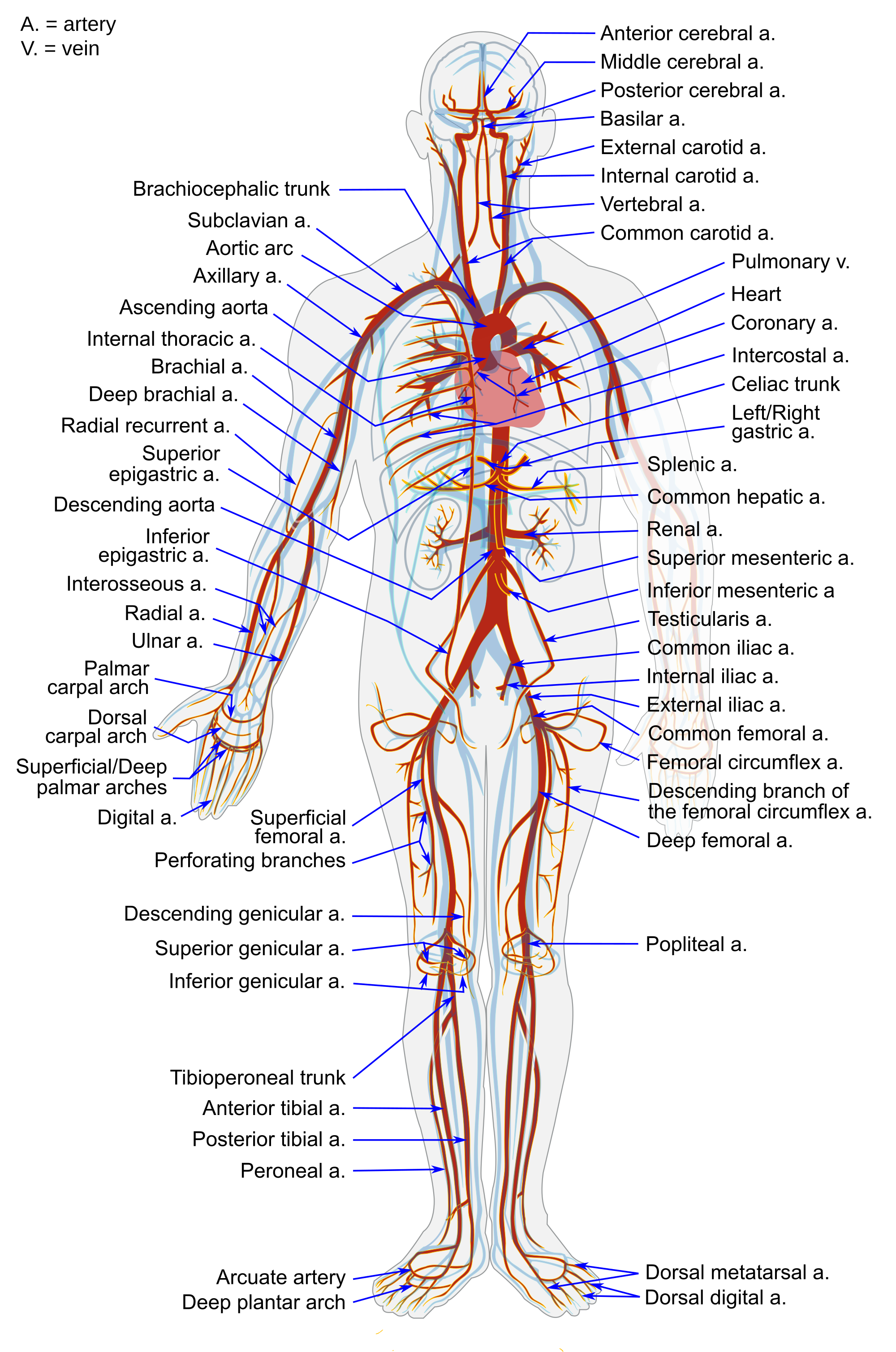
The largest artery in the body is the aorta, which is connected to the heart and extends down into the abdomen (see Figure 14.4.2). The aorta has high-pressure, oxygenated blood pumped directly into it from the left ventricle of the heart. The aorta has many branches, and the branches subdivide repeatedly, with the subdivisions growing smaller and smaller in diameter. The smallest arteries are called arterioles.
Veins
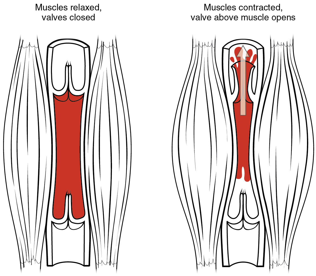
Veins are defined as blood vessels that carry blood toward the heart. Blood traveling through veins is not under pressure from the beating heart. It gets help moving along by the squeezing action of skeletal muscles, for example, when you walk or breathe. It is also prevented from flowing backward by valves in the larger veins, as illustrated in Figure 14.4.3. and as seen in the ultrasonography image in Figure 14.4.4. Veins are called capacitance blood vessels, because the majority of the body’s total volume of blood (about 60 per cent) is contained within veins.
Most veins carry deoxygenated blood, but there are a few exceptions, including the four pulmonary veins. These veins carry oxygenated blood from the lungs to the heart, which then pumps the blood to the rest of the body. In virtually all other veins, hemoglobin is relatively unsaturated with oxygen (about 75 per cent).
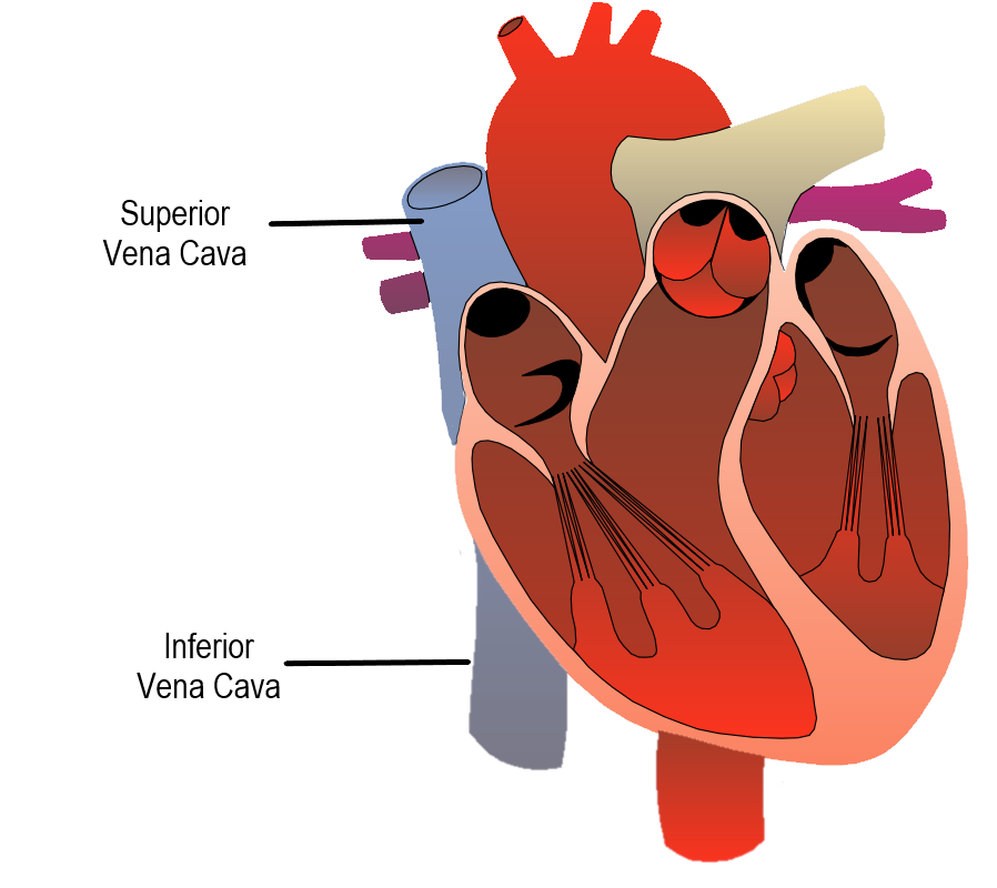
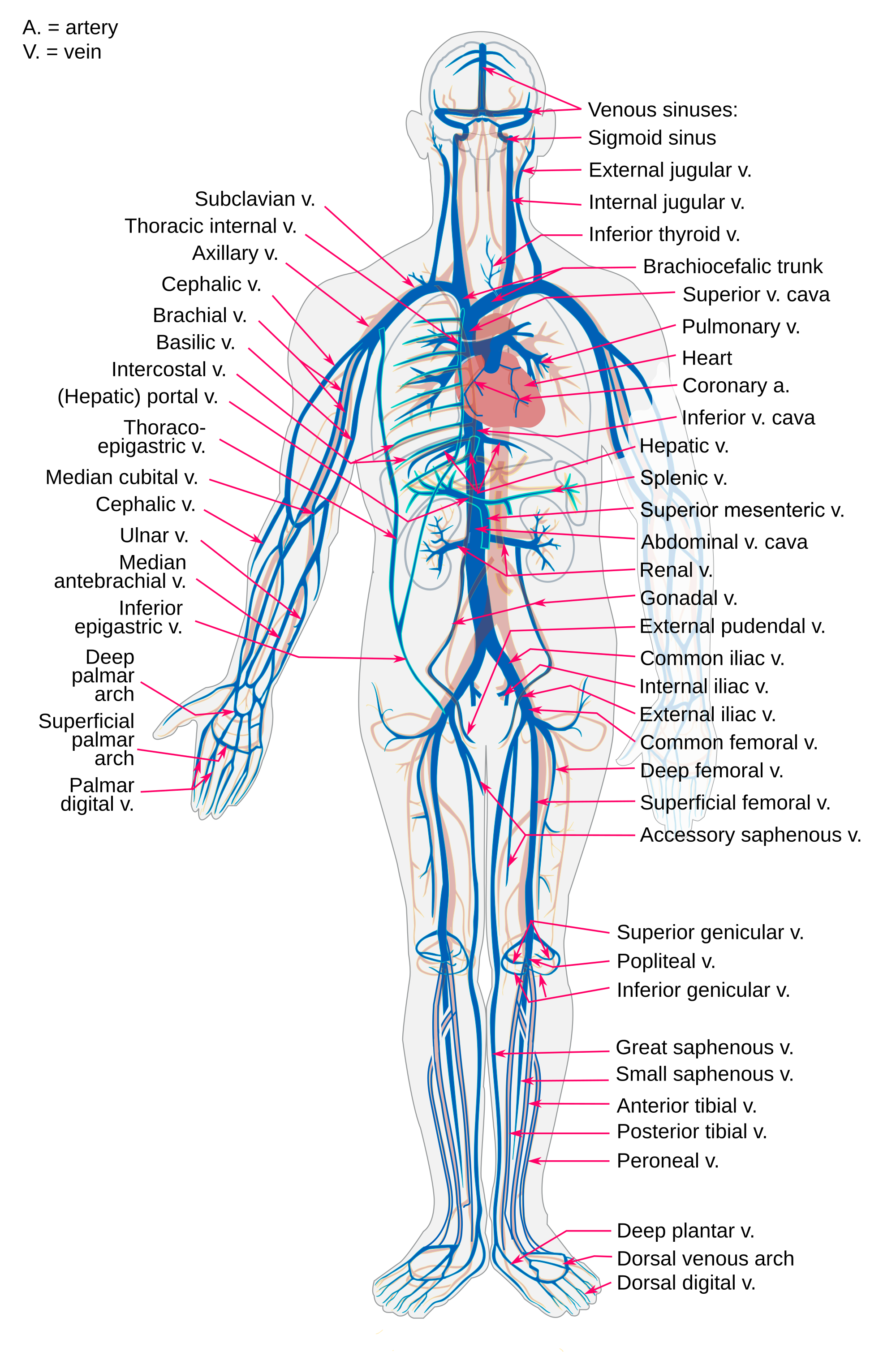
The two largest veins in the body are the superior vena cava — which carries blood from the upper body directly to the right atrium of the heart — and the inferior vena cava, which carries blood from the lower body directly to the right atrium (shown in Figure 14.4.5). Like arteries, veins form a complex, branching system of larger and smaller vessels. The smallest veins are called venules. They receive blood from capillaries and transport it to larger veins. Each venule receives blood from multiple capillaries. See the major veins of the human body in Figure 14.4.6.
Capillaries
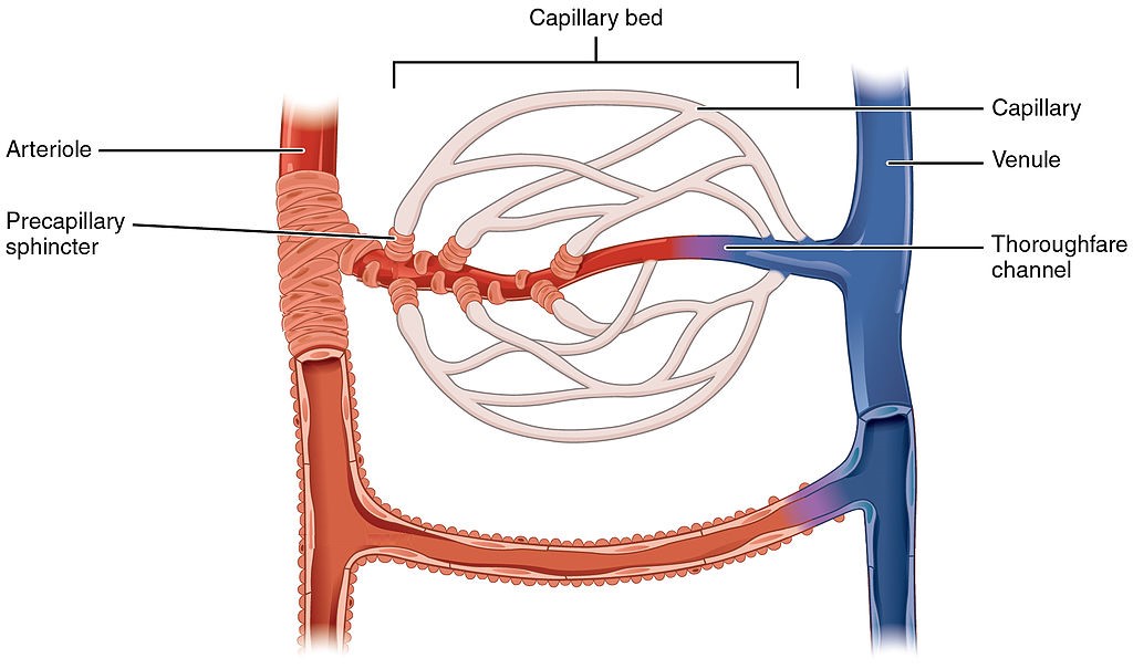
Capillaries are the smallest blood vessels in the cardiovascular system. They are so small that only one red blood cell at a time can squeeze through a capillary, and then only if the red blood cell deforms. Capillaries connect arterioles and venules, as shown in Figure 14.4.7. Capillaries generally form a branching network of vessels, called a capillary bed, that provides a large surface area for the exchange of substances between the blood and surrounding tissues.
Structure of Blood Vessels
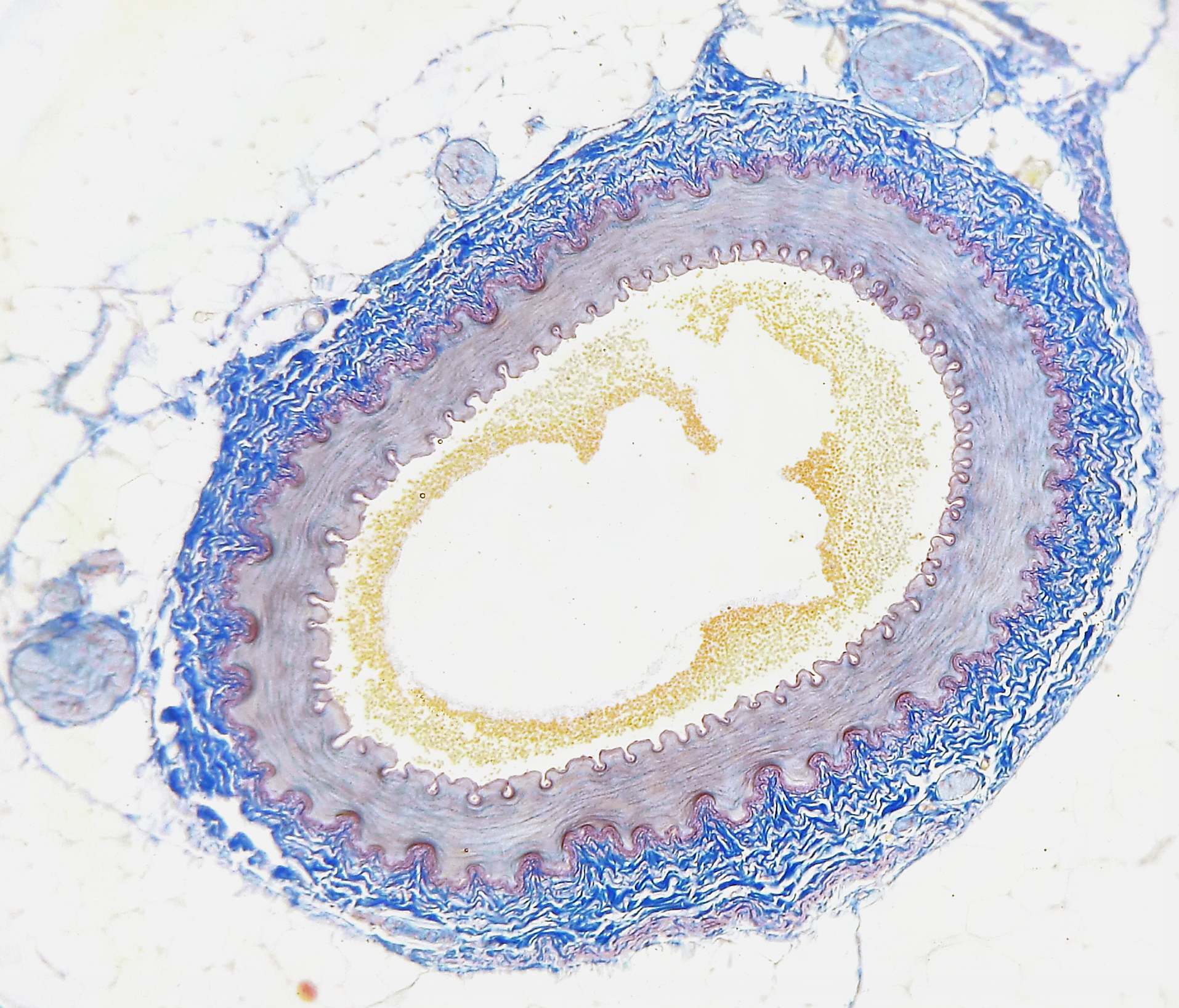
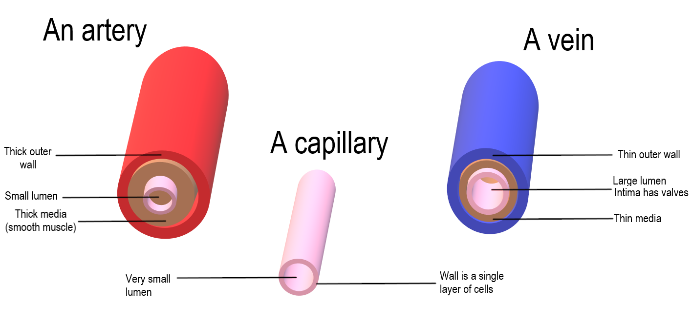
All blood vessels are basically hollow tubes with an internal space, called a lumen, through which blood flows. The lumen of an artery is shown in cross section in the photomicrograph (Figure 14.4.8). The width of blood vessels varies, but they all have a lumen. The walls of blood vessels differ depending on the type of vessel. In general, arteries and veins are more similar to one another than to capillaries in the structure of their walls.
Walls of Arteries and Veins
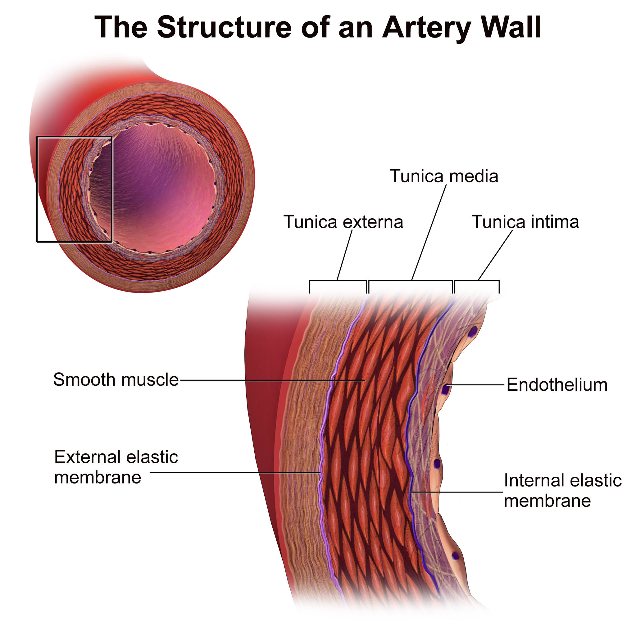
The walls of both arteries and veins have three layers: the tunica intima, tunica media, and tunica adventitia. You can see the three layers for an artery in the Figure 14.4.9.
- The tunica intima is the inner layer of arteries and veins. It is also the thinnest layer, consisting of a single layer of endothelial cells surrounded by a thin layer of connective tissues. It reduces friction between the blood and the inside of the blood vessel walls.
- The tunica media is the middle layer of arteries and veins. In arteries, this is the thickest layer. It consists mainly of elastic fibres and connective tissues. In arteries, this is the thickest layer, because it also contains smooth muscle tissues, which control the diameter of the vessels- as such, the width of the tunic media can be helpful in distinguishing arteries from veins.
- The tunica externa (also called tunica adventitia) is the outer layer of arteries and veins. It consists of connective tissue, and also contains nerves. In veins, this is the thickest layer. In general, the tunica externa protects and strengthens vessels, and attaches them to surrounding structures.
Capillary Walls
The walls of capillaries consist of little more than a single layer of epithelial cells. Being just one cell thick, the walls are well-suited for the exchange of substances between the blood inside them and the cells of surrounding tissues. Substances including water, oxygen, glucose, and other nutrients, as well as waste products (such as carbon dioxide), can pass quickly and easily through the extremely thin walls of capillaries. See figure 14.4.9 for a comparison of the structure of arteries, veins and capillaries.
Blood Pressure
The blood in arteries is normally under pressure because of the beating of the heart. The pressure is highest when the heart contracts and pumps out blood, and lowest when the heart relaxes and refills with blood. (You can feel this variation in pressure in your wrist or neck when you count your pulse.) Blood pressure is a measure of the force that blood exerts on the walls of arteries. It is generally measured in millimetres of mercury (mm Hg), and expressed as a double number — a higher number for systolic pressure when the ventricles contract, and a lower number for diastolic pressure when the ventricles relax. Normal blood pressure is generally defined as less than 120 mm Hg (systolic)/80 mm Hg (diastolic) when measured in the arm at the level of the heart. It decreases as blood flows farther away from the heart and into smaller arteries.
As arteries grow smaller, there is increasing resistance to blood flow through them, because of the blood’s friction against the arterial walls. This resistance restricts blood flow so that less blood reaches smaller, downstream vessels, thus reducing blood pressure before the blood flows into the tiniest vessels, the capillaries. Without this reduction in blood pressure, capillaries would not be able to withstand the pressure of the blood without bursting. By the time blood flows through the veins, it is under very little pressure. The pressure of blood against the walls of veins is always about the same — normally no more than 10 mm Hg.
Vasoconstriction and Vasodilation
Smooth muscles in the walls of arteries can contract or relax to cause vasoconstriction (narrowing of the lumen of blood vessels) or vasodilation (widening of the lumen of blood vessels). This allows the arteries — especially the arterioles — to contract or relax as needed to help regulate blood pressure. In this regard, the arterioles act like an adjustable nozzle on a garden hose. When they narrow, the increased friction with the arterial walls causes less blood to flow downstream from the narrowing, resulting in a drop in blood pressure. These actions are controlled by the autonomic nervous system in response to pressure-sensitive sensory receptors in the walls of larger arteries.
Arteries can also dilate or constrict to help regulate body temperature, by allowing more or less blood to flow from the warm body core to the body’s surface. In addition, vasoconstriction and vasodilation play roles in the fight-or-flight response, under control of the sympathetic nervous system. Vasodilation allows more blood to flow to skeletal muscles, and vasoconstriction reduces blood flow to digestive organs.
Feature: My Human Body
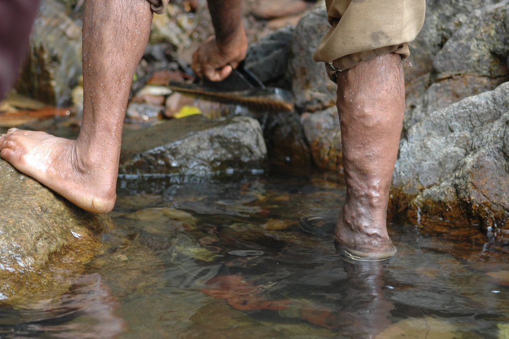
The lumpy appearance of this man’s leg (Figure 14.4.10) is caused by varicose veins. Do you have varicose veins? If you do, you may wonder whether they are a sign of a significant health problem. You may also wonder whether you should have them treated, and if so, what treatments are available. As is usually the case, when it comes to your health, knowledge is power.
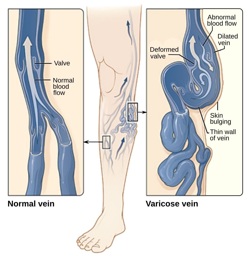
Varicose veins are veins that have become enlarged and twisted, because their valves have become ineffective (see Figure 14.4.11). As a result, blood pools in the veins and stretches them out. Varicose veins occur most frequently in the superficial veins of the legs, but they may also occur in other parts of the body. They are most common in older adults, females, and people who have a family history of the condition. Obesity and pregnancy also increase the risk of developing varicose veins. A job that requires standing for long periods of time, chronic constipation, and long-term alcohol consumption are additional risk factors.
Varicose veins usually are not serious. For many people, they are only a cosmetic issue. In severe cases, however, varicose veins may cause pain and other problems. The affected leg(s) may feel heavy and achy, especially after long periods of standing. Ankles may become swollen by the end of the day. Minor injuries may bleed more than normal. The skin over the varicosity may become red, dry, and itchy. In very severe cases, skin ulcers may develop.
If you are concerned about varicose veins, call them to the attention of your doctor, who can determine the best course of action for your case. There are many potential treatments for varicose veins. Some of the treatments have potential adverse side effects, and with many of the treatments, varicose veins may return. The best treatment for a given patient depends in part on the severity of the condition.
- If varicose veins are not serious, conservative treatment options may be recommended. These include avoiding standing or sitting for long periods, frequently elevating the legs, and wearing graduated compression stockings.
- For more serious cases, less conservative, but non-surgical options may be advised. These include sclerotherapy, in which medicine is injected into the veins to make them shrink. Another non-surgical approach is endovenous thermal ablation. In this type of treatment, laser light, radio-frequency energy, or steam is used to heat the walls of the veins, causing them to shrink and collapse.
- For the most serious cases, surgery may be the best option. The most invasive surgery is vein stripping, in which all or part of the main trunk of a vein is tied off and removed from the leg while the patient is under general anesthesia. In a less invasive surgery, called ambulatory phlebectomy, short segments of a vein are removed through tiny incisions under local anesthesia.
14.4 Summary
- Blood vessels are the part of the cardiovascular system that carries blood throughout the human body. They are long, hollow,tube-like structures. There are three major types of blood vessels: arteries, veins, and capillaries.
- Arteries are blood vessels that carry blood away from the heart. Most arteries carry oxygenated blood. The largest artery is the aorta, which is connected to the heart and extends into the abdomen. Blood moves through arteries due to pressure from the beating of the heart.
- Veins are blood vessels that carry blood toward the heart. Most veins carry deoxygenated blood. The largest veins are the superior vena cava and inferior vena cava. Blood moves through veins by the squeezing action of surrounding skeletal muscles. Valves in veins prevent backflow of blood.
- Capillaries are the smallest blood vessels. They connect arterioles and venules. They form capillary beds, where substances are exchanged between the blood and surrounding tissues.
- The walls of arteries and veins have three layers. The middle layer is thickest in arteries, in which it contains smooth muscle tissue that controls the diameter of the vessels. The outer layer is thickest in veins, and consists mainly of connective tissue. The walls of capillaries consist of little more than a single layer of epithelial cells.
- Blood pressure is a measure of the force that blood exerts on the walls of arteries. It is expressed as a double number, with the higher number representing systolic pressure when the ventricles contract, and the lower number representing diastolic pressure when the ventricles relax. Normal blood pressure is generally defined as a pressure of less than 120/80 mm Hg.
- Vasoconstriction (narrowing) and vasodilation (widening) of arteries can occur to help regulate blood pressure or body temperature, or change blood flow as part of the fight-or-flight response.
14.4 Review Questions
- What are blood vessels? Name the three major types of blood vessels.
-
- Compare and contrast how blood moves through arteries and veins.
- What are capillaries, and what is their function?
- Does the blood in most veins have any oxygen at all? Explain your answer.
- Explain why it is important that the walls of capillaries are very thin.
14.4 Explore More
How blood pressure works – Wilfred Manzano, TED-Ed, 2015.
What are Varicose Veins? Cleveland Clinic, 2019.
Arteries vs Veins ( Circulatory System ), MooMooMath and Science, 2018.
Attributions
Figure 14.4.1
bodybuilding_PNG24 from pngimg.com is used under a CC BY-NC 4.0 (https://creativecommons.org/licenses/by-nc/4.0/) license.
Figure 14.4.2
Arterial_System_en.svg by Mariana Ruiz Villarreal [LadyofHats] on Wikimedia Commons is in the public domain (https://en.wikipedia.org/wiki/Public_domain).
Figure 14.4.3
Skeletal_Muscle_Vein_Pump by OpenStax College on Wikimedia Commons is used under a CC BY 3.0 (https://creativecommons.org/licenses/by/3.0) license.
Figure 14.4.4
Venous_valve_00013 by Nevit Dilmen on Wikimedia Commons is used under a CC BY-SA 3.0 (https://creativecommons.org/licenses/by-sa/3.0) license.
Figure 14.4.5
Superior and Inferior Vena Cava by ArtFavor (acquired from OCAL) from Freestockphotos.biz, is used under a CC0 1.0 Universal public domain dedication license (https://creativecommons.org/publicdomain/zero/1.0/). Work adapted by Christine Miller.
Figure 14.4.6
Venous_system_en.svg by Mariana Ruiz Villarreal [LadyofHats] on Wikimedia Commons is in the public domain (https://en.wikipedia.org/wiki/Public_domain).
Figure 14.4.7
1024px-2105_Capillary_Bed by OpenStax College on Wikimedia Commons is used under a CC BY 3.0 (https://creativecommons.org/licenses/by/3.0) license.
Figure 14.4.8
Artery by Lord of Konrad on Wikimedia Commons is used under a CC0 1.0 Universal public domain dedication license (https://creativecommons.org/publicdomain/zero/1.0/).
Figure 14.4.9
Blausen_0055_ArteryWallStructure by BruceBlaus on Wikimedia Commons is used under a CC BY 3.0 (https://creativecommons.org/licenses/by/3.0) license.
Figure 14.4.10
Artery Vein Capillary Comparison by Christinelmiller on Wikimedia Commons is used under a CC BY-SA 4.0 (https://creativecommons.org/licenses/by-sa/4.0) license.
Figure 14.4.11
Varicose-veins by Jackerhack at English Wikipedia on Wikimedia Commons is used under a CC BY-SA 2.5 (https://creativecommons.org/licenses/by-sa/2.5) license.
Figure 14.4.12
Varicose_veins-en.svg by Jmarchn on Wikimedia Commons is used under a CC BY-SA 3.0 (https://creativecommons.org/licenses/by-sa/3.0) license. [Work modified from Varicose veins.jpg on Wikimedia Commons from National Heart Lung and Blood Institute (NIH)]
References
Betts, J. G., Young, K.A., Wise, J.A., Johnson, E., Poe, B., Kruse, D.H., Korol, O., Johnson, J.E., Womble, M., DeSaix, P. (2013, June 19). Figure 20.6 Capillary bed [digital image]. In Anatomy and Physiology (Section 20.1). OpenStax. https://openstax.org/books/anatomy-and-physiology/pages/20-1-structure-and-function-of-blood-vessels
Betts, J. G., Young, K.A., Wise, J.A., Johnson, E., Poe, B., Kruse, D.H., Korol, O., Johnson, J.E., Womble, M., DeSaix, P. (2013, June 19). Figure 20.15 Skeletal muscle pump [digital image]. In Anatomy and Physiology (Section 20.2). OpenStax. https://openstax.org/books/anatomy-and-physiology/pages/20-2-blood-flow-blood-pressure-and-resistance
Blausen.com Staff. (2014). Medical gallery of Blausen Medical 2014. WikiJournal of Medicine 1 (2). DOI:10.15347/wjm/2014.010. ISSN 2002-4436.
Cleveland Clinic. (2019, December 30). What are varicose veins? YouTube. https://www.youtube.com/watch?v=9Wf8bLXVwFI&feature=youtu.be
MooMooMath and Science. (2018, April 5). Arteries vs veins ( Circulatory System ). YouTube. https://www.youtube.com/watch?v=hnjMdXSyA5o&feature=youtu.be
TED-Ed. (2015, July 23). How blood pressure works – Wilfred Manzano. YouTube. https://www.youtube.com/watch?v=Ab9OZsDECZw&feature=youtu.be
A hollow, tube-like structure through which blood flows in the cardiovascular system; vein, artery, or capillary.
Refers to the body system consisting of the heart, blood vessels and the blood. Blood contains oxygen and other nutrients which your body needs to survive. The body takes these essential nutrients from the blood.
A type of blood vessel that carries blood away from the heart and toward the lungs or body.
The main artery of the body, supplying oxygenated blood to the circulatory system. In humans it passes over the heart from the left ventricle and runs down in front of the backbone.
A type of blood vessel that carries blood toward the heart from the lungs or body.
A large vein carrying deoxygenated blood into the heart. There are two in humans, the inferior vena cava (carrying blood from the lower body) and the superior vena cava (carrying blood from the head, arms, and upper body).
The smallest type of blood vessel that connects arterioles and venules and that transfers substances between blood and tissues.
The innermost layer of an artery or vein. It is made up of one layer of endothelial cells and is supported by an internal elastic lamina. The endothelial cells are in direct contact with the blood flow.
The middle layer of an artery or vein. It lies between the tunica intima on the inside and the tunica externa on the outside. It consists of connective tissues, elastic fibers and smooth muscle.
The outermost layer of a blood vessel, surrounding the tunica media. It is mainly composed of collagen and, in arteries, is supported by external elastic lamina.
The measure of the force exerted by circulating blood on the walls of arteries.
A narrowing of blood vessels so less blood can flow through them.
The widening of blood vessels. It results from relaxation of smooth muscle cells within the vessel walls, in particular in the large veins, large arteries, and smaller arterioles. The process is the opposite of vasoconstriction, which is the narrowing of blood vessels.
An involuntary human body response mediated by the nervous and endocrine systems that prepares the body to fight or flee from perceived danger.
The division of the autonomic nervous system that controls the fight-or-flight response.
