
38 4.12 Cell Cycle and Cell Division
Created by: CK-12/Adapted by Christine Miller
So Many Cells!
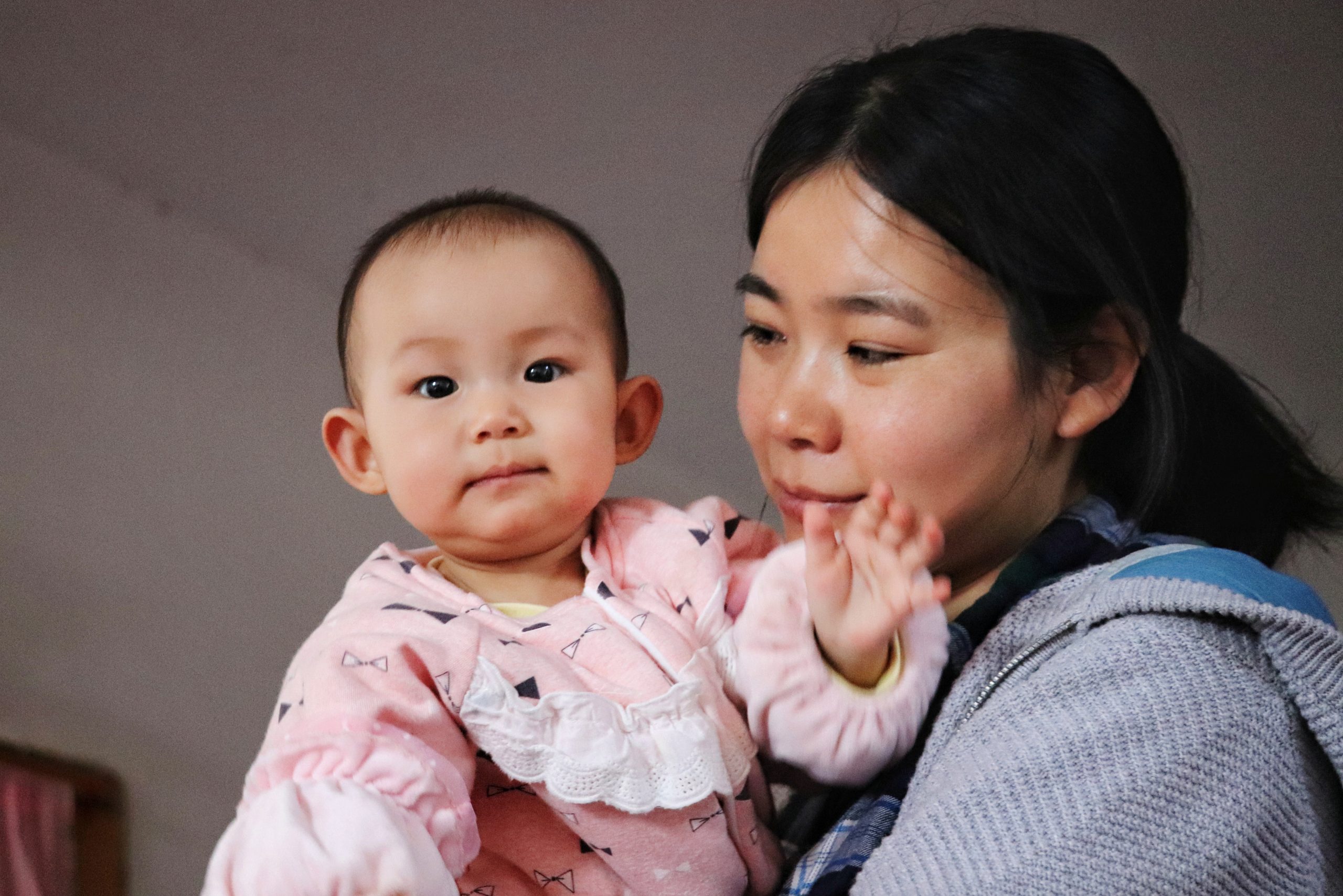
This baby girl (Figure 4.12.1) has a lot of growing to do before she’s as big as her mom. Most of her growth will be the result of cell division. By the time she is an adult, her body will consist of trillions of cells. Cell division is just one of the stages that all cells go through during their life. This includes cells that are harmful, such as cancer cells. Cancer cells divide more often than normal cells, causing them to grow out of control. In fact, this is how cancer cells cause illness. In this concept, you will read about how cells divide, what other stages cells go through, and what causes cancer cells to divide out of control and harm the body.
The Cell Cycle
Cell division is just one of several stages that a cell goes through during its lifetime. The cell cycle is a repeating series of events that includes growth, DNA synthesis, and cell division. The cell cycle in prokaryotes is quite simple: the cell grows, its DNA replicates, and the cell divides. In eukaryotes, the cell cycle is more complicated.
Eukaryotic Cell Cycle
The diagram in Figure 4.12.2 represents the cell cycle of a eukaryotic cell. As you can see, the eukaryotic cell cycle has several phases. The mitotic phase (M) actually includes both mitosis and cytokinesis. This is when the nucleus and then the cytoplasm divide. The other three phases (G1, S, and G2) are generally grouped together as interphase. During interphase, the cell grows, performs routine life processes, and prepares to divide. These phases are discussed below.
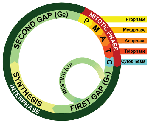
Interphase
The interphase of the eukaryotic cell cycle can be subdivided into the three phases described below, which are represented in Figure 4.12.2.
- Growth Phase 1 (G1): During this phase, the cell grows rapidly, while performing routine metabolic processes. It also makes proteins needed for DNA replication and copies some of its organelles in preparation for cell division. A cell typically spends most of its life in this phase. This phase is also known as gap phase 1.
- Synthesis Phase (S): During this phase, the cell’s DNA is copied in the process of DNA replication, in order to prepare for the upcoming mitotic phase.
- Growth Phase 2 (G2): During this phase, the cell makes final preparations to divide. For example, it makes additional proteins and organelles. This phase is also known as gap phase 2.
Control of the Cell Cycle
If the cell cycle occurred without regulation, cells might go from one phase to the next before they were ready. What controls the cell cycle? How does the cell know when to grow, synthesize DNA, and divide? The cell cycle is controlled mainly by regulatory proteins. These proteins control the cycle by signaling the cell to either start or delay the next phase of the cycle. They ensure that the cell completes the previous phase before moving on. Regulatory proteins control the cell cycle at key checkpoints, which are shown in Figure 4.12.3. There are a number of main checkpoints.
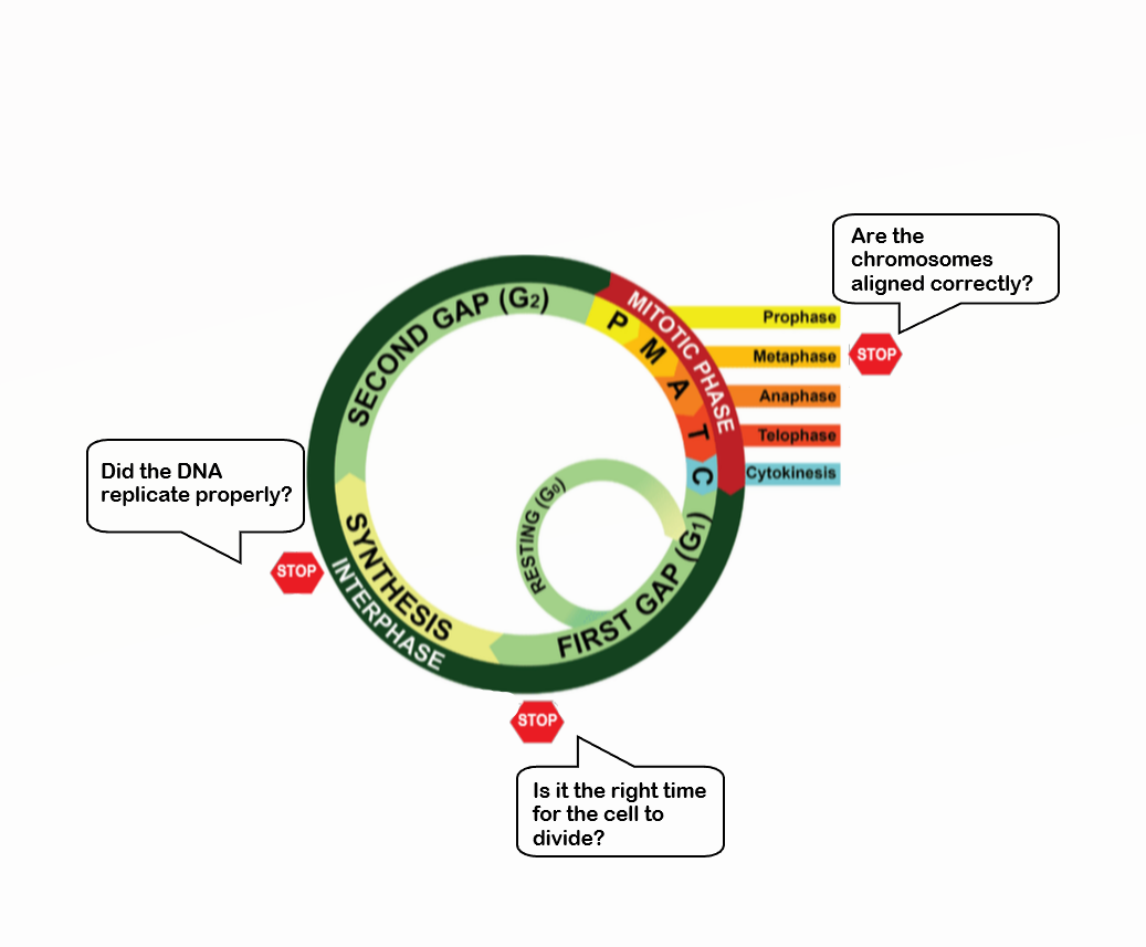
Checkpoints in the eukaryotic cell cycle ensure that the cell is ready to proceed before it moves on to the next phase of the cycle.
- The G1 checkpoint, just before entry into S phase, makes the key decision of whether the cell should divide.
- The S checkpoint determines if the DNA has been replicated properly.
- The mitosis checkpoint ensures that all the chromosomes are properly aligned before the cell is allowed to divide.
Cancer and the Cell Cycle
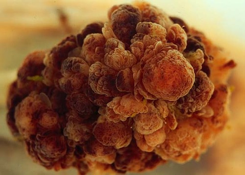
Cancer is a disease that occurs when the cell cycle is no longer regulated. This happens because a cell’s DNA becomes damaged. Damage can occur due to exposure to hazards, such as radiation or toxic chemicals. Cancerous cells generally divide much faster than normal cells. which may end up forming a mass of abnormal cells called a tumor (see Figure 4.12.4). The rapidly dividing cells take up nutrients and space that normal cells need. This can damage tissues and organs and eventually lead to death.
Cell Division
Cell division is the process in which one cell, called the parent cell, divides to form two new cells, referred to as daughter cells. How this happens depends on whether the cell is prokaryotic or eukaryotic. Cell division is simpler in prokaryotes than eukaryotes because prokaryotic cells themselves are simpler. Prokaryotic cells have a single circular chromosome, no nucleus, and few other organelles. Eukaryotic cells, in contrast, have multiple chromosomes contained within a nucleus and many other organelles. All of these cell parts must be duplicated and separated when the cell divides.
Before a eukaryotic cell divides, all of the DNA in the cell’s multiple chromosomes is replicated. Its organelles are also duplicated. Cell division occurs in two major steps, called mitosis and cytokinesis, both of which are described in greater detail in Chapter 5.
- The first step in the division of a eukaryotic cell is mitosis, a multi-phase process in which the nucleus of the cell divides. During mitosis, the nuclear envelope (membrane) breaks down and later reforms. The chromosomes are also sorted and separated to ensure that each daughter cell receives a complete set of chromosomes.
- The second major step is cytokinesis. This step, which also occurs in prokaryotic cells, is when the cytoplasm divides, forming two daughter cells.
Feature: Human Biology in the News
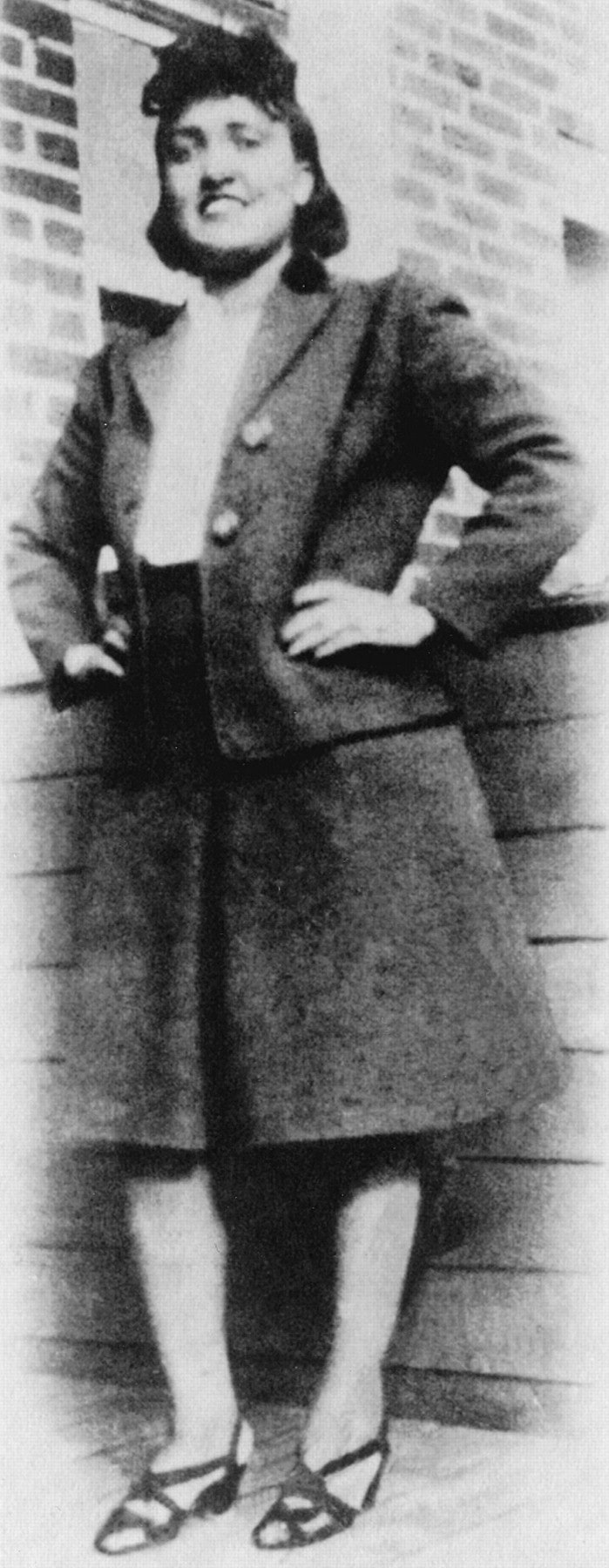
Henrietta Lacks sought treatment for her cancer at Johns Hopkins University Hospital at a time when researchers were trying to grow human cells in the lab for medical testing. Despite many attempts, the cells always died before they had undergone many cell divisions. Mrs. Lacks’s doctor, Howard Jones, took a small sample of cells from her tumor without her knowledge and gave them to a Johns Hopkins researcher, George Gey, who tried to grow them on a culture plate. For the first time in history, human cells grown on a culture plate kept dividing… and dividing and dividing and dividing. Copies of Henrietta’s cells — called HeLa cells, for her name (Henrietta Lacks) — are still alive today. In fact, there are currently billions of HeLa cells in laboratories around the world!
Why Henrietta’s cells lived on when other human cells did not is still something of a mystery, but they are clearly extremely hardy and resilient cells. By 1953, when researchers learned of their ability to keep dividing indefinitely, factories were set up to start producing the cells commercially on a large scale for medical research. Since then, HeLa cells have been used in thousands of studies and have made possible hundreds of medical advances. Jonas Salk, for example, used the cells in the early 1950s to test his polio vaccine. Over the decades since then, HeLa cells have been used to make important discoveries in the study of cancer, AIDS, and many other diseases. The cells were even sent to space on early space missions to learn how human cells respond to zero gravity. HeLa cells were also the first human cells ever cloned, and their genes were some of the first ever mapped. It is almost impossible to overestimate the profound importance of HeLa cells to human biology and medicine.
You would think that Henrietta’s name would be well known in medical history for her unparalleled contributions to biomedical research. However, until 2010, her story was virtually unknown. That year, a science writer named Rebecca Skloot published a nonfiction book, The Immortal Life of Henrietta Lacks. Based on a decade of research, this riveting account became an almost instantaneous best seller. As of 2016, Oprah Winfrey and collaborators planned to make a movie based on the book, and in recent years, numerous articles about Henrietta Lacks have appeared in the press.
Ironically, Henrietta herself never knew her cells had been taken, and neither did her family. While her cells were making a lot of money and building scientific careers, her children were living in poverty, too poor to afford medical insurance. The story of Henrietta Lacks and her immortal cells raises ethical issues about human tissues and who controls them in biomedical research. There is no question that Henrietta Lacks deserves far more recognition for her contribution to the advancement of science and medicine.
If you want to learn more about Henrietta Lacks and her immortal cells, read Rebecca Skloot’s The Immortal Life of Henrietta Lacks (or watch the movie, if it is available). You can also watch the short video below about Henrietta Lacks and her immortal cells by Robin Bulleri:
The immortal cells of Henrietta Lacks – Robin Bulleri, TED-Ed, 2016.
4.12 Summary
- The cell cycle is a repeating series of events that includes growth, DNA synthesis, and cell division. The cycle is more complicated in eukaryotic than prokaryotic cells.
- In a eukaryotic cell, the cell cycle has two major phases: mitotic phase and interphase. During mitotic phase, first the nucleus and then the cytoplasm divide. During interphase, the cell grows, performs routine life processes, and prepares to divide.
- The cell cycle is controlled mainly by regulatory proteins that signal the cell to either start or delay the next phase of the cycle. They ensure that the cell completes the previous phase before moving on. There are a number of main checkpoints in the regulation of the cell cycle.
- Cancer is a disease that occurs when the cell cycle is no longer regulated, often because the cell’s DNA has become damaged. Cancerous cells grow out of control and may form a mass of abnormal cells called a tumor.
- The cell division phase of the cell cycle in a eukaryotic cell occurs in two major steps: mitosis — when the nucleus divides — and cytokinesis, when the cytoplasm divides and two daughter cells form.
4.12 Review Questions
-
- Explain why cell division is more complex in eukaryotic than prokaryotic cells.
- Using a technique called flow cytometry, scientists can distinguish between cells with the normal amount of DNA and those that contain twice the normal amount of DNA as they go through the cell cycle. Which phases of the cell cycle will have cells with twice the amount of DNA? Explain your answer.
- What were scientists trying to do when they took tumor cells from Henrietta Lacks? Why did they specifically use tumor cells to try to achieve their goal?
4.12 Explore More
The Cell Cycle (and cancer) [Updated], The Amoeba Sisters, 2018.
Attributions
Figure 4.12.1
Mom and baby by Taiying Lu on Unsplash is used under the Unsplash License (https://unsplash.com/license).
Figure 4.12.2
Cell Cycle by LadyofHats; CK-12 Foundation is used under a CC BY-NC 3.0 (https://creativecommons.org/licenses/by-nc/3.0/) license.
![]() ©CK-12 Foundation Licensed under
©CK-12 Foundation Licensed under ![]() • Terms of Use • Attribution
• Terms of Use • Attribution
Figure 4.12.3
Cell Cycle Checkpoints by LadyofHats; CK-12 Foundation is used and adapted by Christine Miller under a CC BY-NC 3.0 (https://creativecommons.org/licenses/by-nc/3.0/) license.
![]() ©CK-12 Foundation Licensed under
©CK-12 Foundation Licensed under ![]() • Terms of Use • Attribution
• Terms of Use • Attribution
Figure 4.12.4
Cancer cells forming a tumour by Ed Uthman, MD on Wikimedia Commons is released into the public domain (https://en.wikipedia.org/wiki/Public_domain).
Figure 4.12.5
Henrietta Lacks by Oregon State University on Flickr is used under a CC BY-SA 2.0 (https://creativecommons.org/licenses/by-sa/2.0/) license.
References
Amoeba Sisters. (2018, March 20). The cell cycle (and cancer) [Updated]. YouTube. https://www.youtube.com/watch?v=QVCjdNxJreE&feature=youtu.be
TED-Ed. (2016, February 8). The immortal cells of Henrietta Lacks – Robin Bulleri. YouTube. https://www.youtube.com/watch?v=22lGbAVWhro&feature=youtu.be
Wikipedia contributors. (2020, June 23). Henrietta Lacks. In Wikipedia. https://en.wikipedia.org/w/index.php?title=Henrietta_Lacks&oldid=964020268
Wikipedia contributors. (2020, May 11). Howard W. Jones. In Wikipedia. https://en.wikipedia.org/w/index.php?title=Howard_W._Jones&oldid=956033806
Wikipedia contributors. (2020, July 1). George Otto Gey. In Wikipedia. https://en.wikipedia.org/w/index.php?title=George_Otto_Gey&oldid=965394045
Wikipedia contributors. (2020, July 6). Johns Hopkins Hospital. In ,Wikipedia. https://en.wikipedia.org/w/index.php?title=Johns_Hopkins_Hospital&oldid=966348552
Wikipedia contributors. (2020, June 28). Jonas Salk. In Wikipedia. https://en.wikipedia.org/w/index.php?title=Jonas_Salk&oldid=964883129
Wikipedia contributors. (2020, April 14). Rebecca Skloot. In Wikipedia. https://en.wikipedia.org/w/index.php?title=Rebecca_Skloot&oldid=950837115
Wikipedia contributors. (2020, February 21). The immortal life of Henrietta Lacks. In Wikipedia. https://en.wikipedia.org/w/index.php?title=The_Immortal_Life_of_Henrietta_Lacks&oldid=941942679
A cycle of growth and division that cells go through. It includes interphase (G1, S, and G2) and the mitotic phase.
Cells which lack membrane-bound structures, specifically a nucleus. Instead they generally have a single circular chromosome located in an area of the cell called the nucleoid.
Cells which have a nucleus enclosed within membranes, unlike prokaryotes, which have no membrane-bound organelles.
The longest stage in the eukaryotic cell cycle during which the cell acquires nutrients, creates and uses proteins and other molecules, and starts the process of cell division by replicating the DNA.
A group of diseases involving abnormal cell growth with the potential to invade or spread to other parts of the body.
Deoxyribonucleic acid - the molecule carrying genetic instructions for the development, functioning, growth and reproduction of all known organisms and many viruses.
A mass of tissue that's formed by an accumulation of abnormal cells.
The process by which a parent cell divides into two or more daughter cells. Cell division usually occurs as part of a larger cell cycle.
A tiny cellular structure that performs specific functions within a cell.
A part of the cell cycle when replicated chromosomes are separated into two new nuclei and then subsequent cell division gives rise to genetically identical cells in which the number of chromosomes is maintained.
A structure made up of two lipid bilayer membranes which in eukaryotic cells surrounds the nucleus, which encases the genetic material. Also know as the nuclear membrane.
A threadlike structure of nucleic acids and protein found in the nucleus of most living cells, carrying genetic information in the form of genes.
The part of the cell division process during which the cytoplasm of a single eukaryotic cell divides into two daughter cells. Cytoplasmic division begins during or after the late stages of nuclear division in mitosis and meiosis.
An immortal cell line used in scientific research. It is the oldest and most commonly used human cell line. The line was derived from cervical cancer cells taken on February 8, 1951 from Henrietta Lacks, a patient who died of cancer on October 4, 1951. The cell line was found to be remarkably durable and prolific, which warrants its extensive use in scientific research.
