
102 11.2 Introduction to the Skeletal System
Created by CK-12 Foundation/Adapted by Christine Miller

Skull and Cross-Bones
The skull and cross-bones symbol has been used for a very long time to represent death, perhaps because after death and decomposition, bones are all that remain. Many people think of bones as dead, dry, and brittle. These adjectives may correctly describe the bones of a preserved skeleton, but the bones of a living human being are very much alive. Living bones are also strong and flexible. Bones are the major organs of the skeletal system.
Overview of the Skeleton System
The skeletal system is the organ system that provides an internal framework for the human body. Why do you need a skeletal system? Try to imagine what you would look like without it. You would be a soft, wobbly pile of skin containing muscles and internal organs, but no bones. You might look something like a very large slug. Not that you would be able to see yourself — folds of skin would droop down over your eyes and block your vision, because of your lack of skull bones. You could push the skin out of the way, if you could only move your arms, but you need bones for that, as well!
Components of the Skeletal System
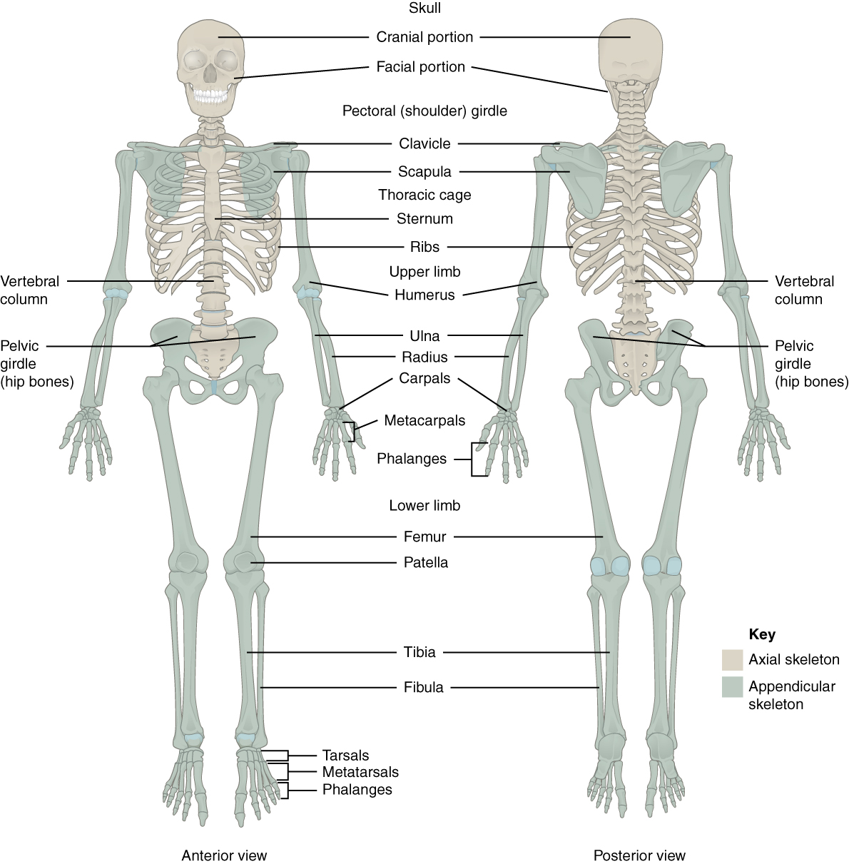
In adults, the skeletal system includes 206 bones, many of which are shown in Figure 10.2.2 below. Bones are organs made of supportive connective tissues, mainly the tough protein collagen. Bones contain blood vessels, nerves, and other tissues, and they are hard and rigid, due to deposits of calcium and other mineral salts within their living tissues. Spots where two or more bones meet are called joints. Many joints allow bones to move like levers. Your elbow, for example, is a joint that allows you to bend and straighten your arm.
Besides bones, the skeletal system includes cartilage and ligaments.
- Cartilage is a type of dense connective tissue, made of tough protein fibres. It is strong, but flexible and very smooth. It covers the ends of bones at joints, providing a smooth surface for bones to move over.
- Ligaments are bands of dense fibrous connective tissue that hold bones together. They keep the bones of the skeleton in place.
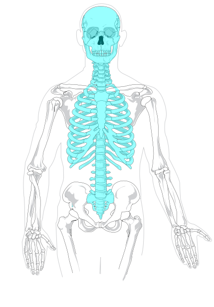
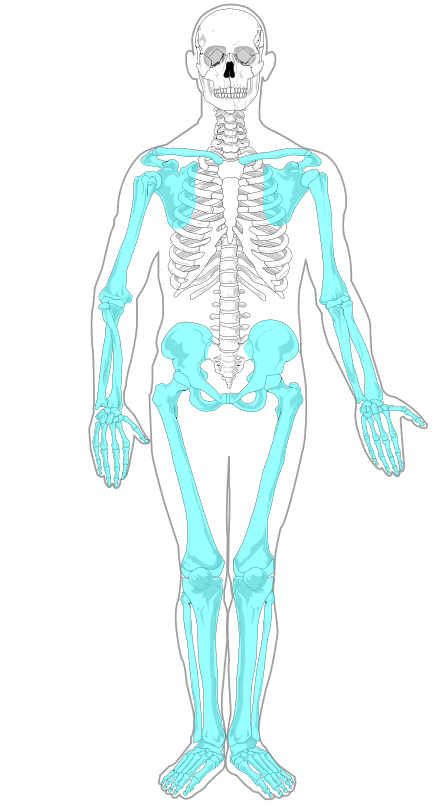
Axial and Appendicular Skeletons
The skeleton is traditionally divided into two major parts: the axial skeleton and the appendicular skeleton, both of which are pictured below (Figure 10.2.3 and Figure 10.2.4 respectively).
- The axial skeleton forms the axis of the body. It includes the skull, vertebral column (spine), and rib cage. The bones of the axial skeleton — along with ligaments and muscles — allow the human body to maintain its upright posture. The axial skeleton also transmits weight from the head, trunk, and upper extremities down the back to the lower extremities. In addition, the bones protect the brain and organs in the chest.
- The appendicular skeleton forms the appendages and their attachments to the axial skeleton. It includes the bones of the arms and legs, hands and feet, and shoulder and pelvic girdles. The bones of the appendicular skeleton make possible locomotion and other movements of the appendages. They also protect the major organs of digestion, excretion, and reproduction.
Functions of the Skeletal System
The skeletal system has many different functions that are necessary for human survival. Some of the functions, such as supporting the body, are relatively obvious. Other functions are less obvious but no less important. Three tiny bones (hammer, anvil, and stirrup) inside the middle ear, for example, transfer sound waves into the inner ear.
Support, Shape, and Protection
The skeleton supports the body and gives it shape. Without the rigid bones of the skeletal system, the human body would be just a bag of soft tissues, as described above. The bones of the skeleton are very hard and provide protection to the delicate tissues of internal organs. For example, the skull encloses and protects the soft tissues of the brain, and the vertebral column protects the nervous tissues of the spinal cord. The vertebral column, ribcage, and sternum (breast bone) protect the heart, lungs, and major blood vessels. Providing protection to these latter internal organs requires the bones to be able to expand and contract. The ribs and the cartilage that connects them to the sternum and vertebrae are capable of small shifts that allow breathing and other internal organ movements.
Movement
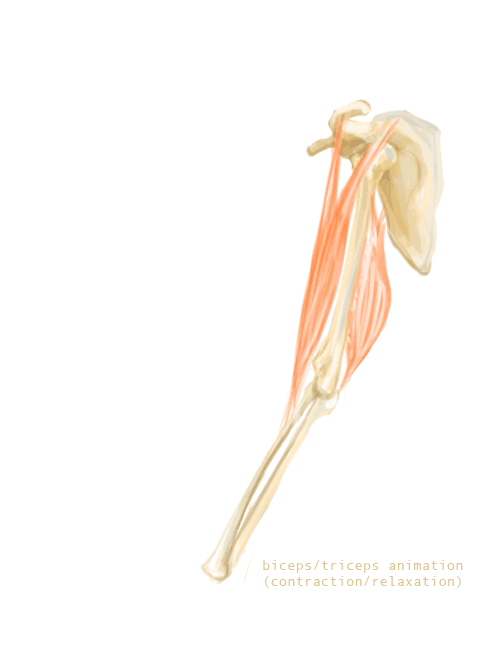
The bones of the skeleton provide attachment surfaces for skeletal muscles. When the muscles contract, they pull on and move the bones. Figure 11.2.5, for example, shows the muscles attached to the bones at the elbow and shoulder. They help stabilize the joint and allow the arm to bend at these two joints. The bones at joints act like levers moving at a fulcrum point, and the muscles attached to the bones apply the force needed for movement.
Hematopoiesis
Hematopoiesis is the process by which blood cells are produced. This process occurs in a tissue called red marrow, which is found inside some bones, including the pelvis, ribs, and vertebrae. Red marrow synthesizes red blood cells, white blood cells, and platelets. Billions of these blood cells are produced inside the bones every day.
Mineral Storage and Homeostasis
Another function of the skeletal system is storing minerals, especially calcium and phosphorus. This storage function is related to the role of bones in maintaining mineral homeostasis. Just the right levels of calcium and other minerals are needed in the blood for normal functioning of the body. When mineral levels in the blood are too high, bones absorb some of the minerals and store them as mineral salts, which is why bones are so hard. When blood levels of minerals are too low, bones release some of the minerals back into the blood. Bone minerals are alkaline (basic), so their release into the blood buffers the blood against excessive acidity (low pH), whereas their absorption back into bones buffers the blood against excessive alkalinity (high pH). In this way, bones help maintain acid-base homeostasis in the blood.
Another way that bones help maintain homeostasis is by acting as an endocrine organ. One endocrine hormone secreted by bone cells is osteocalcin, which helps regulate blood glucose and fat deposition. It increases insulin secretion, as well as cell’s sensitivity to insulin. In addition, it boosts the number of insulin-producing cells and reduces fat stores.
Sexual Dimorphism of the Human Skeleton
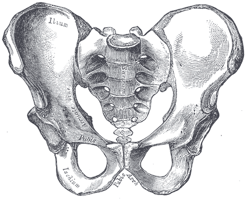
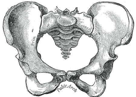
The human skeleton is not as sexually dimorphic as that of many other primate species, although human female skeletons tend to be smaller and less robust than human male skeletons within a given population. There are also subtle differences between males and females in the morphology of the skull, teeth, longs bones, and pelvis. The greatest difference is in the pelvis, because the female pelvis is adapted for child birth. Take a look at the pelvises in Figure 11.2.6 and 11.2.7. How are they different?
 |
 |
11.2 Summary
- The skeletal system is the organ system that provides an internal framework for the human body. In adults, the skeletal system contains 206 bones.
- Bones are organs made of supportive connective tissues, mainly the tough protein collagen. Bones also contain blood vessels, nerves, and other tissues. Bones are hard and rigid, due to deposits of calcium and other mineral salts within their living tissues. Besides bones, the skeletal system includes cartilage and ligaments.
- The skeleton is traditionally divided into two major parts: the axial skeleton (which includes the skull, spine, and rib cage) and the appendicular skeleton (which includes the appendages and the girdles that attach them to the axial skeleton).
- The skeletal system has many different functions, including supporting the body and giving it shape, protecting internal organs, providing attachment surfaces for skeletal muscles, allowing body movements, producing blood cells, storing minerals, helping to maintain mineral homeostasis, and producing endocrine hormones.
- There is relatively little sexual dimorphism in the human skeleton, although the female skeleton tends to be smaller and less robust than the male skeleton. The greatest sex difference is in the pelvis, which is adapted for childbirth in females.
11.2 Review Questions
- What is the skeletal system? How many bones are there in the adult skeleton?
- Describe the composition of bones.
- Besides bones, what other organs are included in the skeletal system?
- Identify the two major divisions of the skeleton.
- List several functions of the skeletal system.
-
- If a person has a problem with blood cell production, what type of bone tissue is most likely involved? Explain your answer.
- What are three forms of homeostasis that the skeletal system regulates? Briefly explain how each one is regulated by the skeletal system.
- What do you think would happen to us if we did not have ligaments? Explain your answer.
- What is a joint? How is cartilage related to joints? Identify one joint in the human body and describe its function.
11.2 Explore More
What can you learn from ancient skeletons? – Farnaz Khatibi, TED-Ed, 2017.
Kathy Reichs on Forensic Anthropology, Cornerstobe Publishing, 2012.
https://www.youtube.com/watch?v=7tKPju8nYi8
Sexual dimorphism in non-human primates – Video Learning – WizScience.com, 2015.
Attributions
Figure 11.2.1
Skull_and_Crossbones.svg by Unknown author on Wikimedia Commons is from The Unicode Standard (this image shows the character U+2620.) All graphic representations of Unicode characters are in the public domain (https://en.wikipedia.org/wiki/Public_domain).
Figure 11.2.2
Skeleton by OpenStax on Wikimedia Commons is used under a CC BY 4.0 (https://creativecommons.org/licenses/by/4.0) license.
Figure 11.2.3
Axial_skeleton_diagram_blank.svg by Quico/ Qllach on Wikimedia Commons is released into the public domain (https://en.wikipedia.org/wiki/Public_domain). (This is a derivative work from Axial skeleton diagram.svg, by Mariana Ruiz Villarreal [LadyofHats].)
Figure 11.2.4
Appendicular_skeleton_diagram_blank.svg by by Quico/ Qllach on Wikimedia Commons is released into the public domain (https://en.wikipedia.org/wiki/Public_domain). (This is a derivative work from Appendicular_skeleton_diagram.svg, by Mariana Ruiz Villarreal [LadyofHats].)
Figure 11.2.5
Animation_triceps_biceps by Niwadare on Wikimedia Commons is used under a CC BY-SA 4.0 (https://creativecommons.org/licenses/by-sa/4.0) license.
Figure 11.2.6
Male pelvisGray241 by Henry Vandyke Carter (1831-1897) on Wikimedia Commons is in the public domain (https://en.wikipedia.org/wiki/Public_domain). (Bartleby.com: Gray’s Anatomy, Plate 241)
Figure 11.2.7
Female pelvisGray242 by Henry Vandyke Carter (1831-1897) on Wikimedia Commons is in the public domain (https://en.wikipedia.org/wiki/Public_domain). (Bartleby.com: Gray’s Anatomy, Plate 242)
Betts, J. G., Young, K.A., Wise, J.A., Johnson, E., Poe, B., Kruse, D.H., Korol, O., Johnson, J.E., Womble, M., DeSaix, P. (2016, May 27). Figure 7.2 Axial and appendicular skeleton [digital image]. In Anatomy and Physiology (Section 7.1). OpenStax. https://openstax.org/books/anatomy-and-physiology/pages/7-1-divisions-of-the-skeletal-system
Cornerstobe Publishing. (2012, November 28). Kathy Reichs on forensic anthropology. YouTube. https://www.youtube.com/watch?v=L101Bvj0lAA
TED-Ed. (2017, June 15). What can you learn from ancient skeletons? – Farnaz Khatibi. YouTube. https://www.youtube.com/watch?v=T24hdchCVIg
VanDyke Carter, H. (1858). Illustration plates 241 and 242. In H. Gray, Anatomy of the Human Body. Lea & Febiger. Bartleby.com, 2000. www.bartleby.com/107/.
Wiz Science. (2015, September 4). Sexual dimorphism in non-human primates – Video Learning – WizScience.com. YouTube. https://www.youtube.com/watch?v=7tKPju8nYi8
The body system composed of bones and cartilage and performs the following critical functions for the human body: supports the body. The skeletal system facilitates movement, protects internal organs, and produces blood cells.
A structure where two or more bones of the skeleton come together.
Supportive connective tissue that provides a smooth surface for the movement of bones at joints. Contains cells called chondrocytes.
A band of dense fibrous connective tissue that holds bones together.
A division of the skeleton that includes the skull, rib cage, and vertebral column.
The bones of the upper and lower limbs, shoulder girdle, and pelvic girdle.
A special sensory organ that collects and amplifies sound waves and information on body position and transforms them into nerve impulses that travel to the brain.
The part of the human skeleton that provides a bony framework for the head and includes bones of the cranium and face.
The central nervous system organ inside the skull that is the control center of the nervous system.
A flexible column of vertebrae that connects the trunk to the skull and encloses the spinal cord; also called spine or backbone.
A specialized tissue found in the central nervous system and the peripheral nervous system. It consists of neurons and supporting cells called neuroglia. The nervous system is responsible for the control of the body and the communication among its parts.
A thin, tubular bundle of central nervous system tissue that extends from the brainstem down the back to the pelvis and connects the brain with the peripheral nervous system.
bony “cage” enclosing the thoracic cavity and consisting of the ribs, thoracic vertebrae, and sternum
A muscular organ in the chest that pumps blood through blood vessels when it contracts.
Two paired organs of the respiratory system in which gas exchange takes place between the blood and the atmosphere.
A hollow, tube-like structure through which blood flows in the cardiovascular system; vein, artery, or capillary.
The process in which red blood cells, white blood cells, and platelets are produced by red bone marrow.
A delicate, highly vascular fibrous tissue containing hematopoietic stem cells. These are blood-forming stem cells.
Paired, fused bones (ilium, pubis, and ischium) that form the hips and attach the legs to the trunk.
One of 33 small bones that make up the vertebral column.
A mineral that is necessary for life. In addition to building bones and keeping them healthy, calcium enables our blood to clot, our muscles to contract, and our heart to beat. About 99% of the calcium in our bodies is in our bones and teeth.
The second most plentiful mineral in your body. The first is calcium. Your body needs phosphorus for many functions, such as filtering waste and repairing tissue and cells.
The ability of an organism to maintain constant internal conditions despite external changes.
An endocrine hormone secreted by bone cells that helps to regulate blood glucose and fat deposition.
Glucose (also called dextrose) is a simple sugar with the molecular formula C6H12O6. Glucose is the most abundant monosaccharide, a subcategory of carbohydrates. Glucose is mainly made by plants and most algae during photosynthesis from water and carbon dioxide, using energy from sunlight.
A hormone made by the pancreas that allows your body to use sugar (glucose) from carbohydrates in the food that you eat for energy or to store glucose for future use.
Differences between the phenotypes of males and females of the same species.
A rigid organ that constitutes part of the vertebrate skeleton in animals.
The main structural protein in the extracellular matrix in the various connective tissues in the body. As the main component of connective tissue, it is the most abundant protein in mammals, making up from 25% to 35% of the whole-body protein content.
