
71 7.4 Tissues
Created by Christine Miller
Figure 7.4.1 Construction — It’s important to have the right materials for the job.
The Right Material for the Job
Building a house is a big job and one that requires a lot of different materials for specific purposes. As you can see in Figure 7.4.1, many different types of materials are used to build a complete house, but each type of material fulfills certain functions. You wouldn’t use insulation to cover your roof, and you wouldn’t use lumber to wire your home. Just as a builder chooses the appropriate materials to build each aspect of a home (wires for electrical, lumber for framing, shingles for roofing), your body uses the right cells for each type of role. When many cells work together to perform a specific function, this is termed a tissue.
Tissues
Groups of connected cells form tissues. The cells in a tissue may all be the same type, or they may be of multiple types. In either case, the cells in the tissue work together to carry out a specific function, and they are always specialized to be able to carry out that function better than any other type of tissue. There are four main types of human tissues: connective, epithelial, muscle, and nervous tissues. We use tissues to build organs and organ systems. The 200 types of cells that the body can produce based on our single set of DNA can create all the types of tissue in the body.
Epithelial Tissue
Epithelial tissue is made up of cells that line inner and outer body surfaces, such as the skin and the inner surface of the digestive tract. Epithelial tissue that lines inner body surfaces and body openings is called mucous membrane. This type of epithelial tissue produces mucus, a slimy substance that coats mucous membranes and traps pathogens, particles, and debris. Epithelial tissue protects the body and its internal organs, secretes substances (such as hormones) in addition to mucus, and absorbs substances (such as nutrients).
The key identifying feature of epithelial tissue is that it contains a free surface and a basement membrane. The free surface is not attached to any other cells and is either open to the outside of the body, or is open to the inside of a hollow organ or body tube. The basement membrane anchors the epithelial tissue to underlying cells.
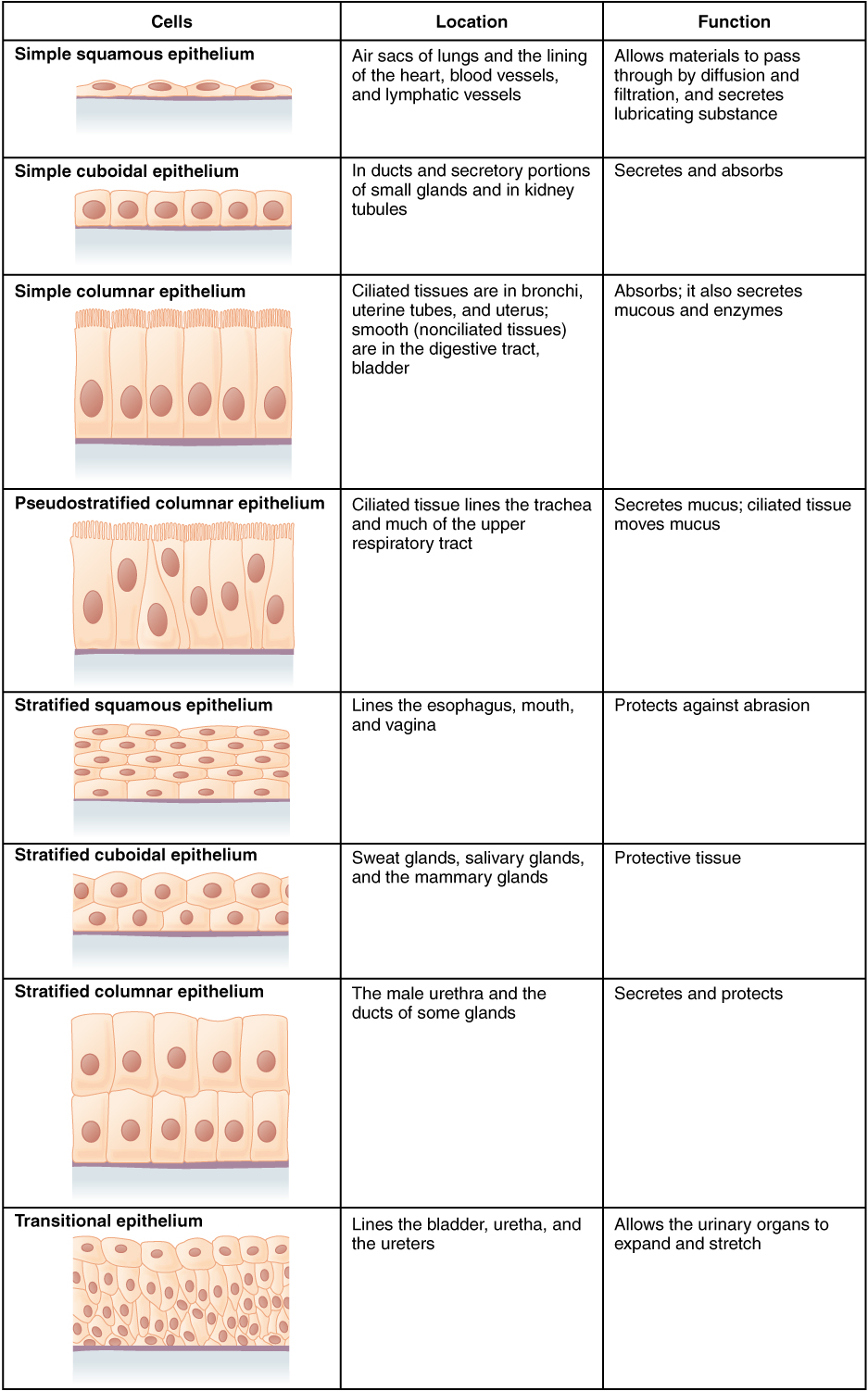
Epithelial tissue is identified and named by shape and layering. Epithelial cells exist in three main shapes: squamous, cuboidal, and columnar. These specifically shaped cells can, depending on function, be layered several different ways: simple, stratified, pseudostratified, and transitional.
Epithelial tissue forms coverings and linings and is responsible for a range of functions including diffusion, absorption, secretion and protection. The shape of an epithelial cell can maximize its ability to perform a certain function. The thinner an epithelial cell is, the easier it is for substances to move through it to carry out diffusion and/or absorption. The larger an epithelial cell is, the more room it has in its cytoplasm to be able to make products for secretion, and the more protection it can provide for underlying tissues. Their are three main shapes of epithelial cells: squamous (which is shaped like a pancake- flat and oval), cuboidal (cube shaped), and columnar (tall and rectangular).
Figure 7.4.2 The shape of epithelial tissues is important.
Epithelial tissue will also organize into different layerings depending on their function. For example, multiple layers of cells provide excellent protection, but would no longer be efficient for diffusion, whereas a single layer would work very well for diffusion, but no longer be as protective; a special type of layering called transitional is needed for organs that stretch, like your bladder. Your tissues exhibit the layering that makes them most efficient for the function they are supposed to perform. There are four main layerings found in epithelial tissue: simple (one layer of cells), stratified (many layers of cells), pseudostratified (appears stratified, but upon closer inspection is actually simple), and transitional (can stretch, going from many layers to fewer layers).
Figure 7.4.3 The layerings found in epithelial tissues is important.
See Table 7.4.1 for a summary of the different layering types and shapes epithelial cells can form and their related functions and locations.
Table 7.4.1
Summary of Epithelial Tissue Cells
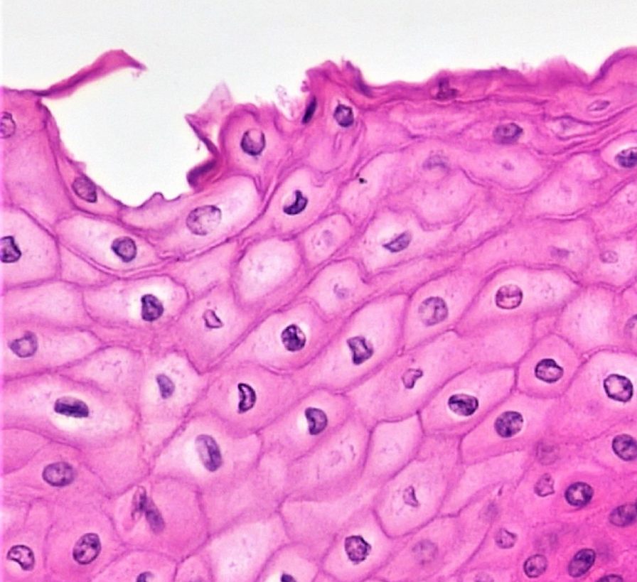
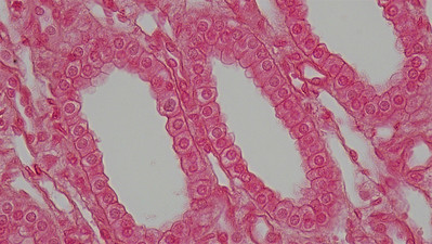
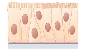
So far, we have identified epithelial tissue based on shape and layering. The representative diagrams we have seen so far are helpful for visualizing the tissue structures, but it is important to look at real examples of these cells. Since cells are too tiny to see with the naked eye, we rely on microscopes to help us study them. Histology is the study of the microscopic anatomy and cells and tissues. See Table 7.4.2 to see some examples of slides of epithelial tissues prepared for the purpose of histology.
Table 7.4.2
Epithelial Tissues and Histological Samples
| Epithelial Tissue Type | Tissue Diagram | Histological Sample |
| Stratified squamous
(from skin) |
 |
|
| Simple cuboidal
(from kidney tubules) |
 |
|
| Pseudostratified ciliated columnar
(from trachea) |
 |
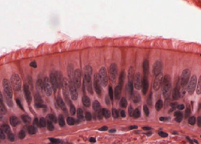 |
Connective Tissue
Bone and blood are examples of connective tissue. Connective tissue is very diverse. In general, it forms a framework and support structure for body tissues and organs. It’s made up of living cells separated by non-living material, called extracellular matrix, which can be solid or liquid. The extracellular matrix of bone, for example, is a rigid mineral framework. The extracellular matrix of blood is liquid plasma.
The key identifying feature of connective tissue is that is is composed of a scattering of cells in a non-cellular matrix. There are three main categories of connective tissue, based on the nature of the matrix. They look very different from one another, which is a reflection of their different functions:
- Fibrous connective tissue: is characterized by a matrix which is flexible and is made of protein fibres including collagen, elastin and possibly reticular fibres. These tissues are found making up tendons, ligaments, and body membranes.
- Supportive connective tissue: is characterized by a solid matrix and is what is used to make bone and cartilage. These tissues are used for support and protection.
- Fluid connective tissue: is characterized by a fluid matrix and includes both blood and lymph.
Fibrous Connective Tissue
Fibrous connective tissue contains cells called fibroblasts. These cells produce fibres of collagen, elastin, or reticular fibre which makes up the matrix of this type of connective tissue. Based on how tightly packed these fibres are and how they are oriented changes the properties, and therefore the function of the fibrous connective tissue.
- Loose fibrous connective tissue: composed of a loose and disorganized weave of collagen and elastin fibres, creating a tissue that is thin and flexible, yet still tough. This tissue, which is also sometimes referred to as “areolar tissue”, is found in membranes and surrounding blood vessels and most body organs. As you can see from the diagram in Figure 7.4.4, loose fibrous connective tissue fulfills the definition of connectives tissue since it is a scattering of cells (fibroblasts) in a non-cellular matrix (a mesh of collagen and elastin fibres). There are two types of specialized loose fibrous connective tissue: reticular and adipose. Adipose tissue stores fat and reticular tissue forms the spleen and lymph nodes.
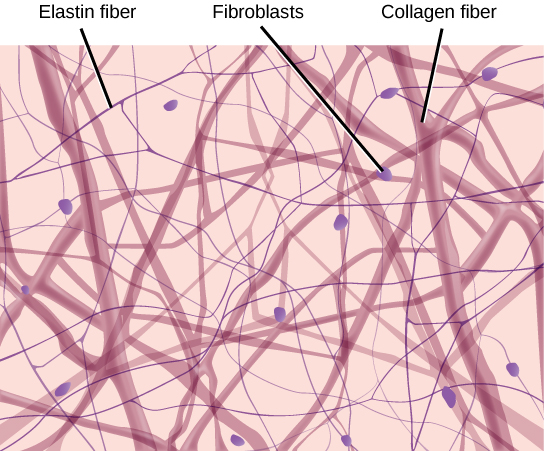
Figure 7.4.4 Diagram of loose fibrous connective tissue consists of a scattering of fibroblasts in a non-cellular matrix of loosely woven collagen and elastin fibres. 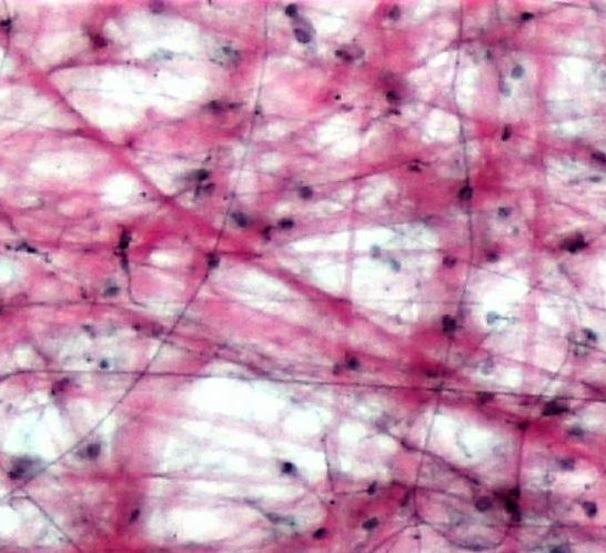
Figure 7.4.5 Microscopic view of loose fibrous connective tissue. - Dense Fibrous Connective Tissue: composed of a dense mat of parallel collagen fibres and a scattering of fibroblasts, creating a tissue that is very strong. Dense fibrous connective tissue forms tendons and ligaments, which connect bones to muscles and/or bones to neighbouring bones.

Figure 7.4.6 Dense fibrous connective tissue is composed of fibroblasts and a dense parallel packing of collagen fibres. 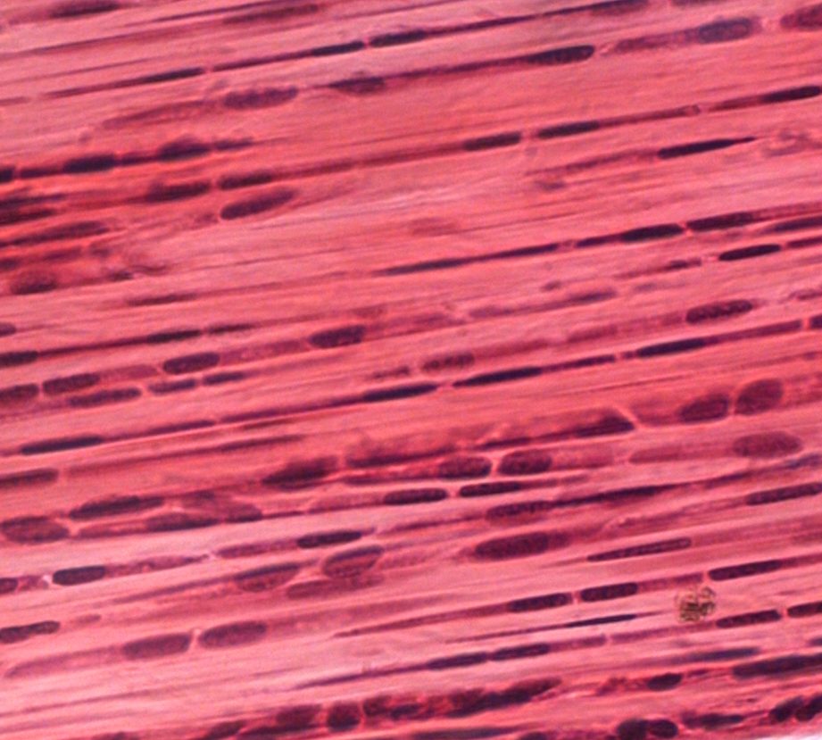
Figure 7.4.7 Microscopic view of dense fibrous connective tissue.
Supportive Connective Tissue
Supportive connective tissue exhibits the defining feature of connective tissue in that it is a scattering of cells in a non-cellular matrix; what sets it apart from other connective tissues is its solid matrix. In this tissue group, the matrix is solid- either bone or cartilage. While fibrous connective tissue contained cells called fibroblasts which produced fibres, supportive connective tissue contains cells that either create bone (osteocytes) or cells that create cartilage (chondrocytes).
Cartilage
Chondrocytes produce the cartilage matrix in which they reside. Cartilage is made up of protein fibres and chondrocytes in lacunae. This is tissue is strong yet flexible and is used many places in the body for protection and support. Cartilage is one of the few tissues that is not vascular (doesn’t have a direct blood supply) meaning it relies on diffusion to obtain nutrients and gases; this is the cause of slow healing rates in injuries involving cartilage. There are three main types of cartilage:
- Hyaline cartilage: a smooth, strong and flexible tissue. Found at the ends of ribs and long bones, in the nose, and comprising the entire fetal skeleton.
- Fibrocartilage: a very strong tissue containing thick bundles of collagen. Found in joints that need cushioning from high impact (knees, jaw).
- Elastic cartilage: contains elastic fibres in addition to collagen, giving support with the benefit of elasticity. Found in earlobes and the epiglottis.
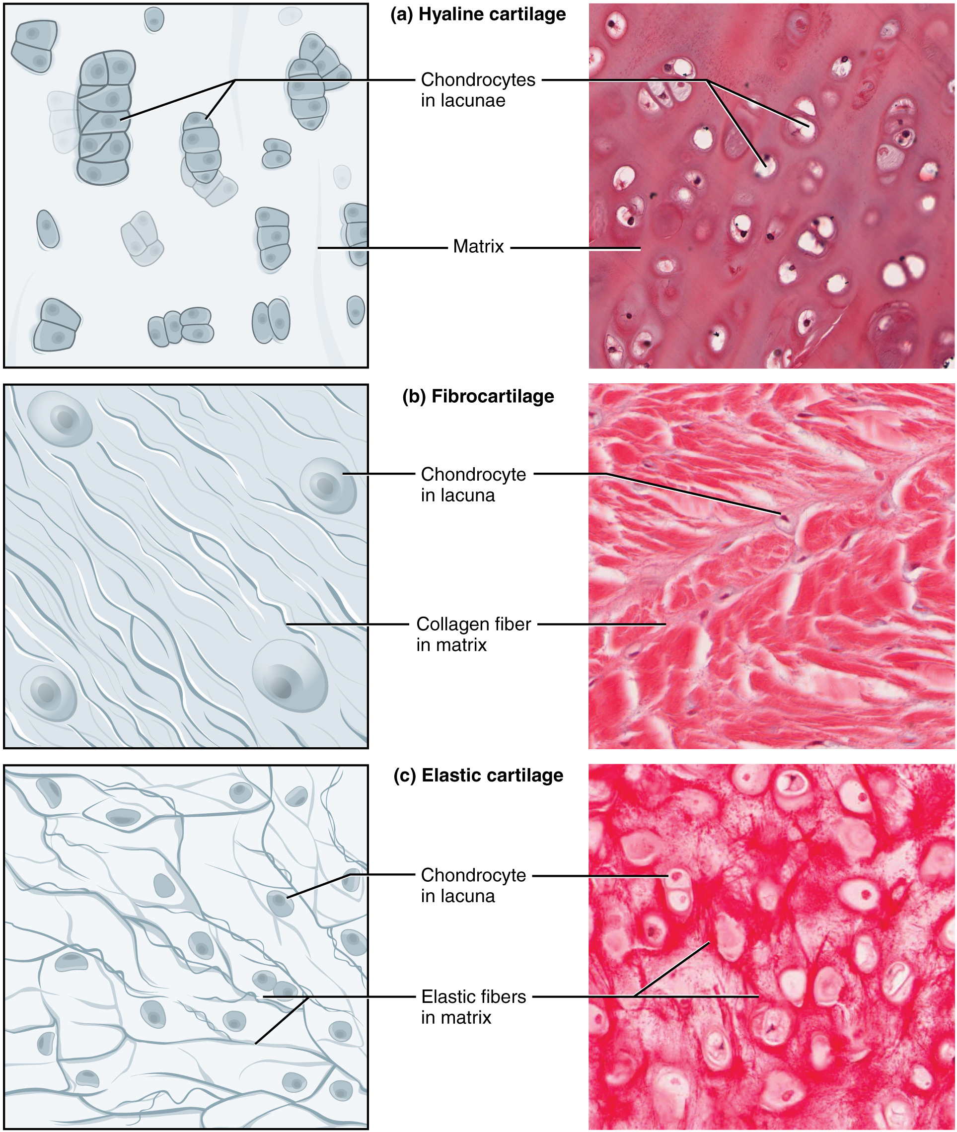
Figure 7.4.8 Three types of cartilage, each with distinct characteristics based on the nature of the matrix.
Bone
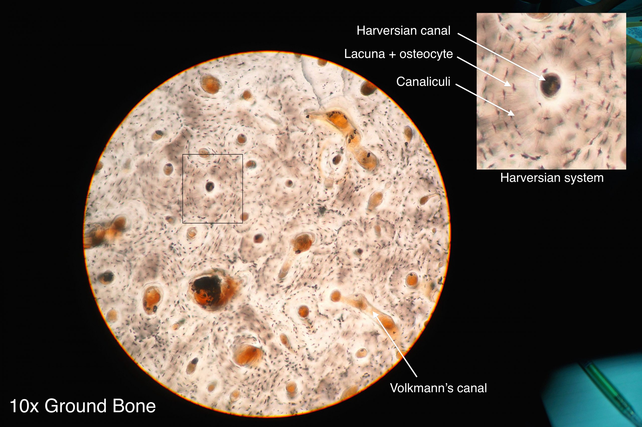
Osteocytes produce the bone matrix in which they reside. Since bone is very solid, these cells reside in small spaces called lacunae. This bone tissue is composed of collagen fibres embedded in calcium phosphate giving it strength without brittleness. There are two types of bone: compact and spongy.
- Compact bone: has a dense matrix organized into cylindrical units called osteons. Each osteon contains a central canal (sometimes called a Harversian Canal) which allows for space for blood vessels and nerves, as well as concentric rings of bone matrix and osteocytes in lacunae, as per the diagram here. Compact bone is found in long bones and forms a shell around spongy bone.
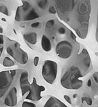
- Spongy bone: a very porous type of bone which most often contains bone marrow. It is found at the end of long bones, and makes up the majority of the ribs, shoulder blades and flat bones of the cranium.
Fluid Connective Tissue
Fluid connective tissue has a matrix that is fluid; unlike the other two categories of connective tissue, the cells that reside in the matrix do not actually produce the matrix. Fibroblasts make the fibrous matrix, chondrocytes make the cartilaginous matrix, osteocytes make the bony matrix, yet blood cells do not make the fluid matrix of either lymph or plasma. This tissue still fits the definition of connective tissue in that it is still a scattering of cells in a non-cellular matrix.
There are two types of fluid connective tissue:
- Blood: blood contains three types of cells suspended in plasma, and is contained in the cardiovascular system.
- Eryththrocytes, more commonly called red blood cells, are present in high numbers (roughly 5 million cells per mL) and are responsible for delivering oxygen from to the lungs to all the other areas of the body. These cells are relatively small in size with a diameter of around 7 micrometres and live no longer than 120 days.
- Leukocytes, often referred to as white blood cells, are present in lower numbers (approximately 5 thousand cells per mL) are responsible for various immune functions. They are typically larger than erythrocytes, but can live much longer, particularly white blood cells responsible for long term immunity. The number of leukocytes in your blood can go up or down based on whether or not you are fighting an infection.
- Thrombocytes, also known as platelets, are very small cells responsible for blood clotting. Thrombocytes are not actually true cells, they are fragments of a much larger cell called a megakaryocyte.
- Lymph: contains a liquid matrix and white blood cells and is contained in the lymphatic system, which ultimately drains into the cardiovascular system.
Figure 7.4.11 A stained lymphocyte surrounded by red blood cells viewed using a light microscope.
Muscular Tissue
Muscular tissue is made up of cells that have the unique ability to contract- which is the defining feature of muscular tissue. There are three major types of muscle tissue, as pictured in Figure 7.4.12 skeletal, smooth, and cardiac muscle tissues.
Skeletal Muscle
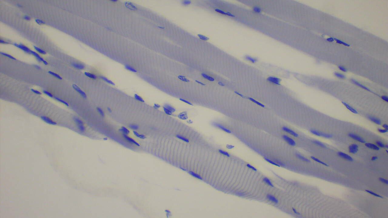
Skeletal muscles are voluntary muscles, meaning that you exercise conscious control over them. Skeletal muscles are attached to bones by tendons, a type of connective tissue. When these muscles shorten to pull on the bones to which they are attached, they enable the body to move. When you are exercising, reading a book, or making dinner, you are using skeletal muscles to move your body to carry out these tasks.
Under the microscope, skeletal muscles are striated (or striped) in appearance, because of their internal structure which contains alternating protein fibres of actin and myosin. Skeletal muscle is described as multinucleated, meaning one “cell” has many nuclei. This is because in utero, individual cells destined to become skeletal muscle fused, forming muscle fibres in a process known as myogenesis. You will learn more about skeletal muscle and how it contracts in the Muscular System.
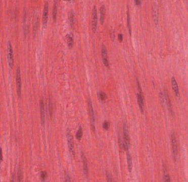
Smooth Muscle
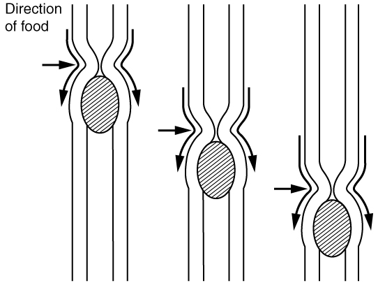
Smooth muscles are nonstriated muscles- they still contain the muscle fibres actin and myosin, but not in the same alternating arrangement seen in skeletal muscle. Smooth muscle is found in the tubes of the body – in the walls of blood vessels and in the reproductive, gastrointestinal, and respiratory tracts. Smooth muscles are not under voluntary control meaning that they operate unconsciously, via the autonomic nervous system. Smooth muscles move substances through a wave of contraction which cascades down the length of a tube, a process termed peristalsis.
Watch the YouTube video “What is Peristalsis” by Mister Science to see peristalsis in action.
What is Peristalsis, Mister Science, 2018.
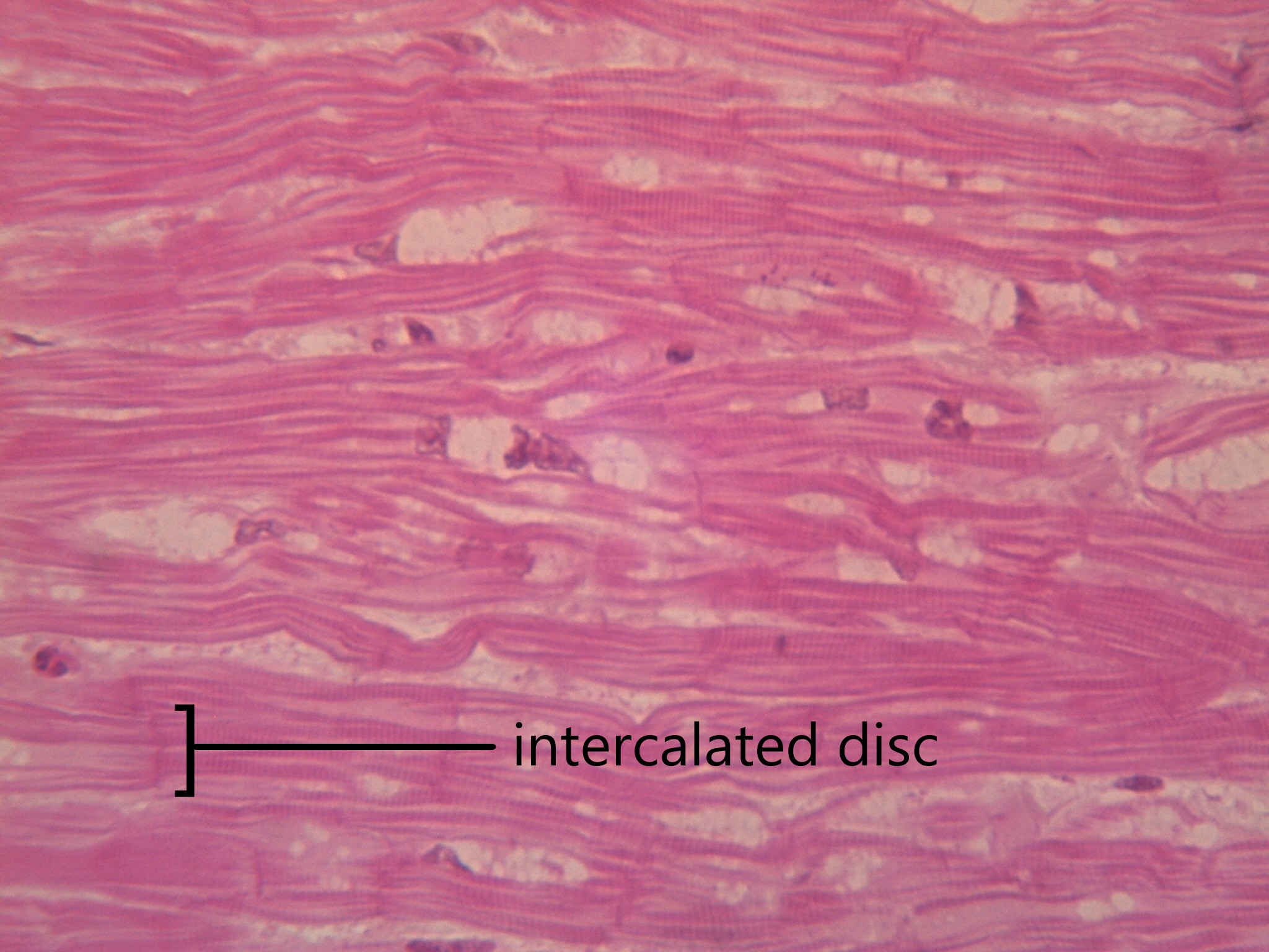
Cardiac Muscle
Cardiac muscles work involuntarily, meaning they are regulated by the autonomic nervous system. This is probably a good thing, since you wouldn’t want to have to consciously concentrate on keeping your heart beating all the time! Cardiac muscle, which is found only in the heart, is mononucleated and striated (due to alternating bands of myosin and actin). Their contractions cause the heart to pump blood. In order to make sure entire sections of the heart contract in unison, cardiac muscle tissue contains special cell junctions called intercalated discs, which conduct the electrical signals used to “tell” the chambers of the heart when to contract.
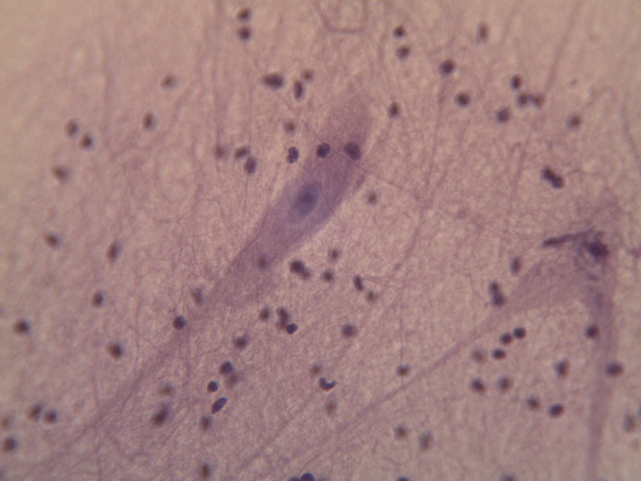
Nervous Tissue
Nervous tissue is made up of neurons and a group of cells called neuroglia (also known as glial cells). Nervous tissue makes up the central nervous system (mainly the brain and spinal cord) and peripheral nervous system (the network of nerves that runs throughout the rest of the body). The defining feature of nervous tissue is that it is specialized to be able to generate and conduct nerve impulses. This function is carried out by neurons, and the purpose of neuroglia is to support neurons.
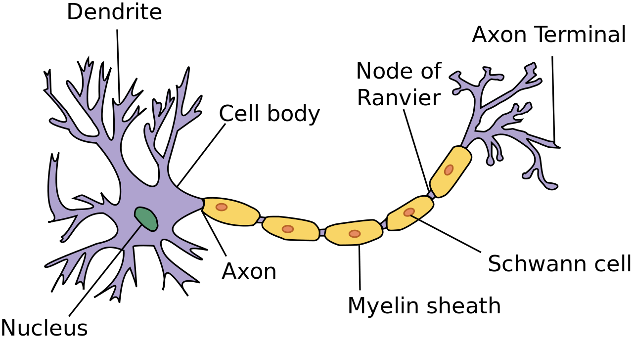
A neuron has several parts to its structure:
- Dendrites which collect incoming nerve impulses
- A cell body, or soma, which contains the majority of the neuron’s organelles, including the nucleus
- An axon, which carries nerve impulses away from the soma, to the next neuron in the chain
- A myelin sheath, which encases the axon and increases that rate at which nerve impulses can be conducted
- Axon terminals, which maintain physical contact with the dendrites of neighbouring neurons
Neuroglia can be understood as support staff for the neuron. The neurons have such an important job, they need cells to bring them nutrients, take away cell waste, and build their mylein sheath. There are many types of neuroglia, which are categorized based on their function and/or their location in the nervous system. Neuroglia outnumber neurons by as much as 50 to 1, and are much smaller. See the diagram in 7.4.17 to compare the size and number of neurons and neuroglia.
Try out this memory game to test your tissues knowledge:
7.4 Summary
- Tissues are made up of cells working together.
- There are four main types of tissues: epithelial, connective, muscular and nervous.
- Epithelial tissue makes up the linings and coverings of the body and is characterized by having a free surface and a basement membrane. Types of epithelial tissue are distinguished by shape of cell (squamous, cuboidal or columnar) and layering (simple, stratified, pseudostratified and transitional). Different epithelial tissues can carry out diffusion, secretion, absorption, and/or protection depending on their particular cell shape and layering.
- Connective tissue provides structure and support for the body and is characterized as a scattering of cells in a non-cellular matrix. There are three main categories of connective tissue, each characterized by a particular type of matrix:
- Fibrous connective tissue contains protein fibres. Both loose and dense fibrous connective tissue belong in this category.
- Supportive connective tissue contains a very solid matrix, and includes both bone and cartilage.
- Fluid connective tissue contains cells in a fluid matrix with the two types of blood and lymph.
- Muscular tissue’s defining feature is that it is contractile. There are three types of muscular tissue: skeletal muscle which is found attached to the skeleton for voluntary movement, smooth muscle which moves substances through body tubes, and cardiac muscle which moves blood through the heart.
- Nervous tissue contains specialized cells called neurons which can conduct electrical impulses. Also found in nervous tissue are neuroglia, which support neurons by providing nutrients, removing wastes, and creating myelin sheath.
7.4 Review Questions
- Define the term tissue.
-
- If a part of the body needed a lining that was both protective, but still able to absorb nutrients, what would be the best type of epithelial tissue to use?
-
- Where do you find skeletal muscle? Smooth muscle? Cardiac muscle?
-
- What are some of the functions of neuroglia?
-
7.4 Explore More
Types of Human Body Tissue, MoomooMath and Science, 2017.
How to 3D print human tissue – Taneka Jones, TED-Ed, 2019.
How bones make blood – Melody Smith, TED-Ed, 2020.
Attributions
Figure 7.4.1
- Construction man kneeling in front of wall by Charles Deluvio on Unsplash is used under the Unsplash License (https://unsplash.com/license).
- Beige wooden frame by Charles Deluvio on Unsplash is used under the Unsplash License (https://unsplash.com/license).
- Tambour on green by Pierre Châtel-Innocention Unsplash is used under the Unsplash License (https://unsplash.com/license).
- Tags: Construction Studs Plumbing Wiring by JWahl on Pixabay is used under the Pixabay License (https://pixabay.com/es/service/license/).
Figure 7.4.2 and Figure 7.4.3
- Simple columnar epithelium tissue by Kamil Danak on Wikimedia Commons is used under a CC BY-SA 3.0 (https://creativecommons.org/licenses/by-sa/3.0/deed.en) license.
- Simple cuboidal epithelium by Kamil Danak on Wikimedia Commons is used under a CC BY-SA 3.0 (https://creativecommons.org/licenses/by-sa/3.0/deed.en) license.
- Simple squamous epithelium by Kamil Danak on Wikimedia Commons is used under a CC BY-SA 3.0 (https://creativecommons.org/licenses/by-sa/3.0/deed.en) license.
Figure 7.4.4
Loose fibrous connective tissue by CNX OpenStax. Biology. on Wikimedial Commons is used under a CC BY 4.0. (https://creativecommons.org/licenses/by/4.0) license.
Figure 7.4.5
Connective Tissue: Loose Aerolar by Berkshire Community College Bioscience Image Library on Flickr is used under a CC0 1.0 Universal public domain dedication (https://creativecommons.org/publicdomain/zero/1.0/) license.
Figure 7.4.6
Dense Fibrous Connective Tissue by by CNX OpenStax. Biology. on Wikimedial Commons is used under a CC BY 4.0. (https://creativecommons.org/licenses/by/4.0) license.
Figure 7.4.7
Dense_connective_tissue-400x by J Jana on Wikimedia Commons is used under a CC BY-SA 4.0 (https://creativecommons.org/licenses/by-sa/4.0/deed.en) license.
Figure 7.4.8
Types_of_Cartilage-new by OpenStax College on Wikipedia Commons is used under a CC BY 3.0 (https://creativecommons.org/licenses/by/3.0) license.
Figure 7.4.9
Compact_bone_histology_2014 by Athikhun.suw on Wikimedia Commons is used under a CC BY-SA 4.0 (https://creativecommons.org/licenses/by-sa/4.0/deed.en) license.
Figure 7.4.10
Bone_normal_and_degraded_micro_structure by Gtirouflet on Wikimedia Commons is used under a CC BY-SA 3.0 (https://creativecommons.org/licenses/by-sa/3.0/deed.en) license.
Figure 7.4.11
Lymphocyte2 by NicolasGrandjean on Wikimedia Commons is used under a CC BY-SA 3.0 (https://creativecommons.org/licenses/by-sa/3.0/deed.en) license. [No machine-readable author provided. NicolasGrandjean is assumed, based on copyright claims.]
Figure 7.4.12
Skeletal_muscle_横纹肌1 by 乌拉跨氪 on Wikimedia Commons is used under a CC BY-SA 4.0 (https://creativecommons.org/licenses/by-sa/4.0/deed.en) license.
Figure 7.4.13
Smooth_Muscle_new by OpenStax on Wikimedia Commons is used under a CC BY 4.0 (https://creativecommons.org/licenses/by/4.0/deed.en) license.
Figure 7.4.14
Peristalsis by OpenStax on Wikimedia Commons is used under a CC BY 3.0 (https://creativecommons.org/licenses/by/3.0/deed.en) license.
Figure 7.4.15
400x Cardiac Muscle by Jessy731 on Flickr is used and adapted by Christine Miller under a CC BY-NC 2.0 (https://creativecommons.org/licenses/by-nc/2.0/) license.
Figure 7.4.16
Neuron.svg by User:Dhp1080 on Wikimedia Commons is used under a CC BY-SA 3.0 (https://creativecommons.org/licenses/by-sa/3.0/deed.en) license.
Figure 7.4.17
400x Nervous Tissue by Jessy731 on Flickr is used under a CC BY-NC 2.0 (https://creativecommons.org/licenses/by-nc/2.0/) license.
Table 7.4.1
Summary of Epithelial Tissue Cells, by OpenStax College on Wikipedia Commons is used under a CC BY 3.0 (https://creativecommons.org/licenses/by/3.0) license.
Table 7.4.2
- Epithelial_Tissues_Stratified_Squamous_Epithelium_(40230842160) by
Berkshire Community College Bioscience Image Library on Wikimedia Commons is used under a CC0 1.0 Universal Public Domain Dedication (https://creativecommons.org/publicdomain/zero/1.0/) license. - Simple cuboidal epithelial tissue histology by Berkshire Community College on Flickr is used under a CC0 1.0 Universal Public Domain Dedication (https://creativecommons.org/publicdomain/zero/1.0/) license.
- Pseudostratified_Epithelium by OpenStax College on Wikimedia Commons is used under a CC BY 3.0 (https://creativecommons.org/licenses/by/3.0) license.
References
Betts, J. G., Young, K.A., Wise, J.A., Johnson, E., Poe, B., Kruse, D.H., Korol, O., Johnson, J.E., Womble, M., DeSaix, P. (2013, April 25). Figure 4.8 Summary of epithelial tissue cells [digital image]. In Anatomy and Physiology (Section 4.2). OpenStax. https://openstax.org/books/anatomy-and-physiology/pages/4-2-epithelial-tissue
Betts, J. G., Young, K.A., Wise, J.A., Johnson, E., Poe, B., Kruse, D.H., Korol, O., Johnson, J.E., Womble, M., DeSaix, P. (2013, April 25). Figure 4.16 Types of cartilage [digital image]. In Anatomy and Physiology (Section 4.3). OpenStax. https://openstax.org/books/anatomy-and-physiology/pages/4-3-connective-tissue-supports-and-protects
Betts, J. G., Young, K.A., Wise, J.A., Johnson, E., Poe, B., Kruse, D.H., Korol, O., Johnson, J.E., Womble, M., DeSaix, P. (2013, April 25). Figure 10.23 Smooth muscle [digital micrograph]. In Anatomy and Physiology (Section 10.8). OpenStax. https://openstax.org/books/anatomy-and-physiology/pages/10-8-smooth-muscle (Micrograph provided by the Regents of University of Michigan Medical School © 2012)
Betts, J. G., Young, K.A., Wise, J.A., Johnson, E., Poe, B., Kruse, D.H., Korol, O., Johnson, J.E., Womble, M., DeSaix, P. (2013, April 25). Figure 22.5 Pseudostratified ciliated columnar epithelium [digital micrograph]. In Anatomy and Physiology (Section 22.1). OpenStax. https://openstax.org/books/anatomy-and-physiology/pages/22-1-organs-and-structures-of-the-respiratory-system
Betts, J. G., Young, K.A., Wise, J.A., Johnson, E., Poe, B., Kruse, D.H., Korol, O., Johnson, J.E., Womble, M., DeSaix, P. (2013, April 25). Figure 23.5 Peristalsis [diagram]. In Anatomy and Physiology (Section 23.2). OpenStax. https://openstax.org/books/anatomy-and-physiology/pages/23-2-digestive-system-processes-and-regulation
Mister Science. (2018). What is peristalsis? YouTube. https://www.youtube.com/watch?v=kVjeNZA5pi4
MoomooMath and Science. (2017, May 18). Types of human body tissue. YouTube. https://www.youtube.com/watch?v=O0ZvbPak4ck&feature=youtu.be
Open Stax. (2016, May 27). Figure 6 Loose connective tissue [digital image]. In OpenStax Biology (Section 33.2). OpenStax CNX. https://cnx.org/contents/GFy_h8cu@10.53:-LfhWRES@4/Animal-Primary-Tissues
Open Stax. (2016, May 27). Figure 7 Fibrous connective tissue from the tendon [digital image]. In OpenStax Biology (Section 33.2). OpenStax CNX. https://cnx.org/contents/GFy_h8cu@10.53:-LfhWRES@4/Animal-Primary-Tissues
TED-Ed. (2019, October 17). How to 3D print human tissue – Taneka Jones. YouTube. https://www.youtube.com/watch?v=uHbn7wLN_3k&feature=youtu.be
TED-Ed. (2020, January 27). How bones make blood – Melody Smith. YouTube. https://www.youtube.com/watch?v=1Qfmkd6C8u8&feature=youtu.be
A cellular organizational level between cells and a complete organ. A tissue is an ensemble of similar cells and their extracellular matrix from the same origin that together carry out a specific function. Organs are then formed by the functional grouping together of multiple tissues.
Tissue which lines the outer surfaces of organs and blood vessels throughout the body, as well as the inner surfaces of cavities in many internal organs. An example is the epidermis, the outermost layer of the skin. There are three principal shapes of epithelial cell: squamous, columnar, and cuboidal.
The study of the microscopic anatomy and cells and tissues.
One of the four basic types of tissue, connective tissue is found in between other tissues everywhere in the body, including the nervous system and generally forms a framework and support structure for body tissues and organs.
A three-dimensional network of extracellular macromolecules, such as collagen, enzymes, and glycoproteins, that provide structural and biochemical support to surrounding cells.
A cell in connective tissue which produces collagen and/or other protein fibers.
a bone cell, formed when an osteoblast becomes embedded in the matrix it has secreted.
A cell which has secreted the matrix of cartilage and become embedded in it.
A small space containing an osteocyte in bone or chondrocyte in cartilage.
A soft tissue that composes muscles in animal bodies, and gives rise to muscles' ability to contract. This is opposed to other components or tissues in muscle such as tendons or perimysium.
A distinctive pattern of smooth muscle contractions that propels foodstuffs distally through the esophagus and intestines.
Unique structural formations found between the myocardial cells of the heart. They play vital roles in bonding cardiac muscle cells together and in transmitting signals between cells.
A specialized tissue found in the central nervous system and the peripheral nervous system. It consists of neurons and supporting cells called neuroglia. The nervous system is responsible for the control of the body and the communication among its parts.

.jpg)