
108 11.8 Case Study Conclusion: A Pain in the Foot
Created by CK-12 Foundation/Adapted by Christine Miller
Case Study Conclusion: A Pain in the Foot
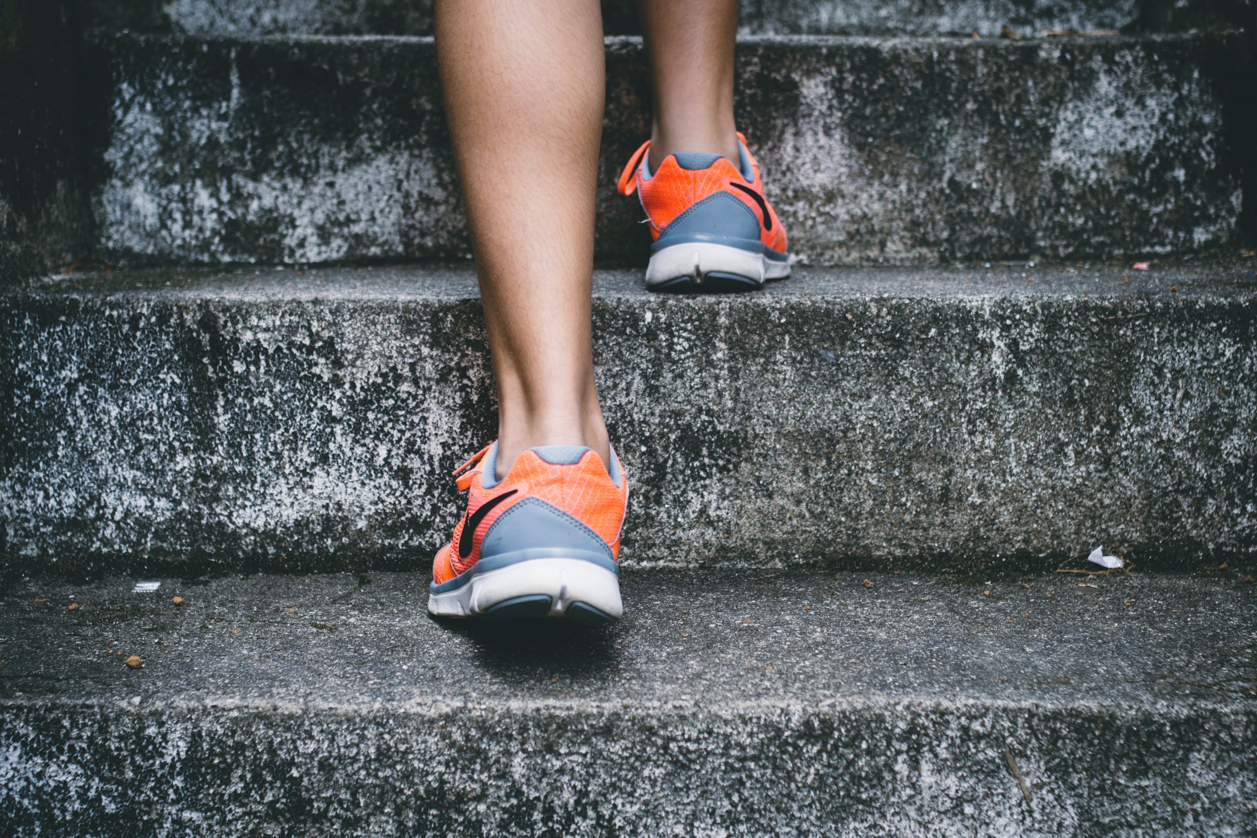
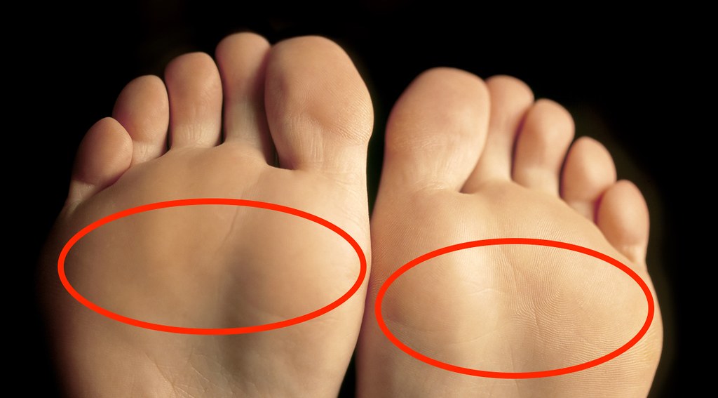
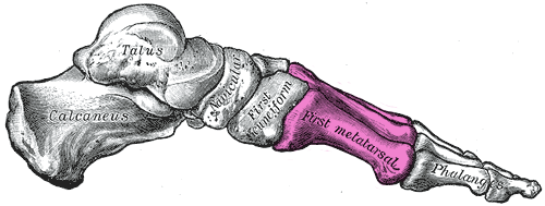
As Sophia discovered in the beginning of the chapter, wearing high heels can result in a condition called metatarsalgia. Metatarsalgia is named for the metatarsal bones, which are the five bones that run through the ball of the foot just behind the toes (highlighted in Figures 11.8.2 and 11.8.3). Wearing high heels causes excessive pressure on the ball of the foot, as described in the beginning of this chapter. Additionally, the toes are forced to pull upward in high heels, which moves the fleshy padding away from the ball of the foot and adds to the overall pressure placed on this region. Over time, this can cause inflammation and direct stress on the bones, resulting in the pain in the ball of the foot known as metatarsalgia. The pain occurs especially in weight-bearing positions, such as standing, walking, or running — which is what Sophia was experiencing. There may also be pain, numbness, or tingling in the toes associated with metatarsalgia.
 |
 |
Wearing high heels can also cause stress fractures in the feet, which are tiny breaks in bone that occur due to repeated mechanical stress. This is caused by the excessive pressure that high heels put on some of the bones of the feet. These fractures are somewhat similar to what occurs in osteoporosis when the bone mass decreases to the point where bones can fracture easily as a person goes about their daily activities. In both cases, a major noticeable injury is not necessary to create the tiny fractures. As you have learned, tiny fractures that accrue over time are the cause of dowager’s hump (or kyphosis), which is often seen in women with osteoporosis.
Don’t think you are immune to stress fractures just because you don’t wear high heels! This injury also commonly occurs in people who participate in sports involving repetitive striking of the foot on the ground, such as running, tennis, basketball, or gymnastics. They may be avoided by taking preventative measures. You should ramp up any increase in activity slowly, cross-train by engaging in a variety of different sports or activities, rest if you experience pain, and wear well-cushioned and supportive running shoes. It is important to know that your cardiovascular and muscular systems adapt to an increase in physical activity much more quickly than the skeletal system.
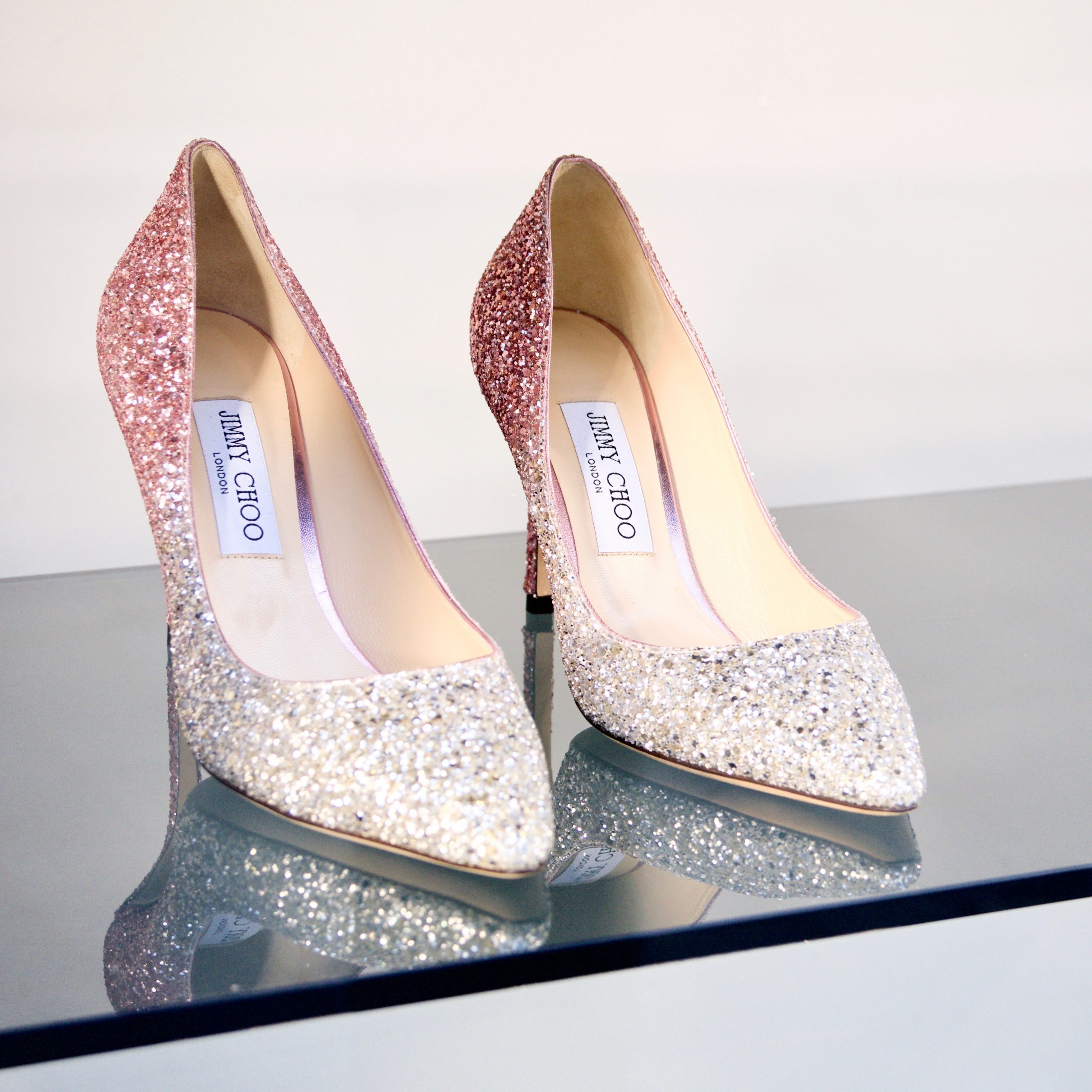
Sophia learned through her online research that wearing high heels can also lead to foot deformities, such as bunions and hammertoes. As you learned in an earlier chapter, a bunion is a protrusion on the side of the foot, most often at the base of the big toe. It can be caused by wearing shoes with a narrow, pointed toe box — a common shape for high heels (see Figure 11.8.4). The pressure of the shoes on the side of the foot causes an enlargement of bone or inflammation of other tissues in the region, which pushes the big toe toward the other toes.
Hammertoes are an abnormal bend in the middle joint of the second, third, or fourth toe (with the big toe being the first toe), causing the toe to be shaped similarly to a hammer. The narrow, pointed toe box of many high heels, combined with the way the toes are squished into the front of the shoe as a result of the height of the heel, can cause the toes to become deformed this way. Treatments for bunions and hammertoe include wearing shoes with a roomy toe box, padding or taping the toes, and toe exercises and stretches. If the bunion or hammertoe does not respond to these treatments, surgery may be necessary to correct the deformity.
Because the bones of the skeleton are connected and work together with other systems to support the body, wearing high heels can also cause physical problems in areas other than the feet. Wearing high heels shifts a person’s posture and alignment, and can put strain on tendons, muscles, and other joints in the body. Research published in 2014 from a team at Stanford University suggests that wearing high heels, particularly if the person is overweight or the heels are very high, may increase the risk of osteoarthritis (OA) in the knee, due to added stress on the knee joint as the person walks. As you have learned, OA results from the breakdown of cartilage and bone at the joint. Because it can only be treated to minimize symptoms — and not for a cure — OA could be an unfortunate long-term consequence of wearing high heels.
Sophia has decided that wearing high heels regularly is not worth the pain and potential long-term damage to her body. After consulting with her doctor, who confirmed she had metatarsalgia, she was able to successfully treat it with ice, rest, and wearing comfortable, supportive shoes instead of heels.
High heels are not the only kind of shoes that can cause problems. Flip-flops, worn-out sneakers, and shoes that are too tight can all cause foot issues. To prevent future problems from her shoe choices, Sophia is following guidelines recommended by medical experts. The guidelines include:
- Wearing shoes that fit well, have plenty of room in the toes, are supportive, and are comfortable right away. There should be no “break-in” period needed for shoes.
- Avoiding high heels, especially those with heels over two inches high, or those that have narrow, pointed toe boxes or very thin heels. The heels pictured in Figure 11.8.4 are an example of a type of shoe that should be avoided.
- If high heels must be worn, it should only be for a limited period of time.
As you have learned in this chapter, your skeletal system carries out a variety of important functions in your body, including physical support. But even though it is strong, your skeletal system can become damaged and deformed — even through such a seemingly innocuous act as wearing a certain type of shoe. Taking good care of your skeletal system is necessary to help it continue to take good care of the rest of you.
Chapter 11 Summary
In this chapter, you learned about the skeletal system. Specifically, you learned that:
- The skeletal system is the organ system that provides an internal framework for the human body. In adults, the skeletal system contains 206 bones.
- Bones are organs made of supportive connective tissues, mainly the tough protein collagen. Bones also contain blood vessels, nerves, and other tissues. Bones are hard and rigid, due to deposits of calcium and other mineral salts within their living tissues. Besides bones, the skeletal system includes cartilage and ligaments.
- The skeletal system has many different functions, including supporting the body and giving it shape, protecting internal organs, providing attachment surfaces for skeletal muscles, allowing body movements, producing blood cells, storing minerals, helping to maintain mineral homeostasis, and producing endocrine hormones.
- There is relatively little sexual dimorphism in the human skeleton, although the female skeleton tends to be smaller and less robust than the male skeleton. The greatest sex difference is in the pelvis, which is adapted for childbirth in females.
- The skeleton is traditionally divided into two major parts: the axial skeleton and the appendicular skeleton.
- The axial skeleton consists of a total of 80 bones. It includes the skull, vertebral column, and rib cage. It also includes the three tiny ossicles in the middle ear and the hyoid bone in the throat.
-
- The skull provides a bony framework for the head. It consists of 22 different bones: eight in the cranium, which encloses the brain, and 14 in the face, which includes the upper and lower jaw.
- The vertebral column is a flexible, S-shaped column of 33 vertebrae that connects the trunk with the skull and encloses the spinal cord. The vertebrae are divided into five regions: cervical, thoracic, lumbar, sacral, and coccygeal regions. The S shape of the vertebral column allows it to absorb shocks and distribute the weight of the body.
- The rib cage holds and protects the organs of the upper part of the trunk, including the heart and lungs. It includes the 12 thoracic vertebrae, the sternum, and 12 pairs of ribs.
- The appendicular skeleton consists of a total of 126 bones. It includes the bones of the four limbs, shoulder girdle, and pelvic girdle. The girdles attach the appendages to the axial skeleton.
-
- Each upper limb consists of 30 bones. There is one bone (called the humerus) in the upper arm, and two bones (called the ulna and radius) in the lower arm. The wrist contains eight carpal bones, the hand contains five metacarpals, and the fingers consist of 14 phalanges. The thumb is opposable to the palm and fingers of the same hand.
- Each lower limb also consists of 30 bones. There is one bone (called the femur) in the upper leg, and two bones (called the tibia and fibula) in the lower leg. The patella covers the knee joint. The ankle contains seven tarsal bones, and the foot contains five metatarsals. The tarsals and metatarsals form the heel and arch of the foot. The bones in the toes consist of 14 phalanges.
- The shoulder girdle attaches the upper limbs to the trunk of the body. It is connected to the axial skeleton only by muscles, allowing mobility of the upper limbs. Bones of the shoulder girdle include a right and left clavicle, and a right and left scapula.
- The pelvic girdle attaches the legs to the trunk of the body and supports the organs of the abdomen. It is connected to the axial skeleton by ligaments. The pelvic girdle consists of two halves that are fused together in adults. Each half consists of three bones: the ilium, pubis, and ischium.
- Bones are organs that consist mainly of bone (or osseous) tissue. Osseous tissue is a type of supportive connective tissue consisting of a collagen matrix that is mineralized with calcium and phosphorus crystals. The combination of flexible collagen and minerals makes bone hard, without making it brittle.
-
- There are two types of osseous tissues: compact bone tissue and spongy bone tissue. Compact bone tissue is smooth and dense. It forms the outer layer of bones. Spongy bone tissue is porous and light, and it is found inside many bones.
- Besides osseous tissues, bones also contain nerves, blood vessels, bone marrow, and periosteum.
- Bone tissue is composed of four different types of bone cells: osteoblasts, osteocytes, osteoclasts, and osteogenic cells. Osteoblasts form new collagen matrix and mineralize it, osteoclasts break down bone, osteocytes regulate the formation and breakdown of bone, and osteogenic cells divide and differentiate to form new osteoblasts. Bone is a very active tissue, constantly being remodeled by the work of osteoblasts and osteoclasts.
- There are six types of bones in the human body: long bones (such as the limb bones), short bones (such as the wrist bones), sesamoid bones (such as the patella), sutural bones in the skull, and irregular bones (such as the vertebrae).
- Early in the development of a human fetus, the skeleton is made almost entirely of cartilage. The relatively soft cartilage gradually turns into hard bone — a process that is called ossification. It begins at a primary ossification center in the middle of bone, and later also occurs at secondary ossification centers in the ends of bone. The bone can no longer grow in length after the areas of ossification meet and fuse at the time of skeletal maturity.
- Throughout life, bone is constantly being replaced in the process of bone remodeling. In this process, osteoclasts resorb bone and osteoblasts make new bone to replace it. Bone remodeling shapes the skeleton, repairs tiny flaws in bones, and helps maintain mineral homeostasis in the blood.
- Bone repair is the natural process in which a bone repairs itself following a bone fracture. This process may take several weeks. In the process, the periosteum produces cells that develop into osteoblasts, and the osteoblasts form new bone matrix to heal the fracture. Bone repair may be affected by diet, age, pre-existing bone disease, or other factors.
- Joints are locations at which bones of the skeleton connect with one another.
- Joints can be classified structurally or functionally, and there is significant overlap between the two types of classifications.
- The structural classification of joints depends on the type of tissue that binds the bones to each other at the joint. There are three types of joints in the structural classification: fibrous, cartilaginous, and synovial joints.
- The functional classification of joints is based on the type and degree of movement that they allow. There are three types of joints in the functional classification: immovable, partly movable, and movable joints.
- A number of disorders affect the skeletal system, including bone fractures and bone cancers. The two most common disorders of the skeletal system are osteoporosis and osteoarthritis.
- Osteoporosis is an age-related disorder in which bones lose mass, weaken, and break more easily than normal bones. The underlying mechanism in all cases of osteoporosis is an imbalance between bone formation and bone resorption in bone remodeling. Osteoporosis may also occur as a side effect of other disorders or certain medications.
-
- Osteoporosis is diagnosed by measuring a patient’s bone density and comparing it with the normal level of peak bone density. Fractures are the most dangerous aspect of osteoporosis. Osteoporosis is rarely fatal, but complications of fractures often are.
- Risk factors for osteoporosis include older age, female sex, European or Asian ancestry, family history of osteoporosis, short stature and small bones, smoking, alcohol consumption, lack of exercise, vitamin D deficiency, poor nutrition, and consumption of soft drinks.
- Osteoporosis is often treated with medications (such as bisphosphonates) that may slow or even reverse bone loss. Preventing osteoporosis includes eliminating any risk factors that can be controlled through changes of behavior, such as undertaking weight-bearing exercise.
- Osteoarthritis (OA) is a joint disease that results from the breakdown of joint cartilage and bone. The most common symptoms are joint pain and stiffness. OA is thought to be caused by mechanical stress on the joints with insufficient self-repair of cartilage, coupled with low-grade inflammation of the joints.
-
- Diagnosis of OA is typically made on the basis of signs and symptoms, such as joint deformities, pain, and stiffness. X-rays or other tests are sometimes used to either support the diagnosis or rule out other disorders. Age is the chief risk factor for OA. Other risk factors include joint injury, excess body weight, and a family history of OA.
- OA cannot be cured, but the symptoms can often be treated successfully. Treatments may include exercise, efforts to decrease stress on joints, pain medications, and surgery to replace affected hip or knee joints.
As you have learned in this chapter, one of the important functions of the skeletal system is to allow movement of the body. But it doesn’t do it alone. Movement is caused by the contraction of muscles, which pull on the bones, causing them to move. Read the next chapter to learn about this and other important functions of the muscular system.
Chapter 11 Review
-
-
- Why does the rib cage need to be flexible? Why can it be flexible?
- In general, what do “girdles” in the skeletal system do?
- Would swimming be more effective as an exercise for preventing osteoporosis or as a treatment for osteoarthritis? Explain your answer.
- Explain why some of the vertebrae become misshapen in the condition called dowager’s hump (or kyphosis).
- Explain why osteoarthritis often involves inflammation in the joints.
- Osteoporosis can involve excess bone resorption, as well as insufficient production of new bone tissue. What are the two main bone cell types that carry out these processes, respectively?
- Describe two roles that calcium in bones play in the body.
-
Attributions
Figure 11.8.1
Running Shoes by bruno-nascimento-PHIgYUGQPvU [photo] by Bruno Nascimento on Unsplash is used under the Unsplash License (https://unsplash.com/license).
Figure 11.8.2
Metatarsalgia/ Best Shoes for Metatarsalgia by Esther Max on Flickr is used under a CC BY 2.0 (https://creativecommons.org/licenses/by/2.0/) license.
Figure 11.8.3
Gray290_-_Mratatarsus (1) by Henry Vandyke Carter (1831-1897) (Revised by Warren H. Lewis, coloured by Was a bee) on Wikimedia Commons is in the public domain (https://en.wikipedia.org/wiki/Public_domain. (Bartleby.com: Gray’s Anatomy, Plate 290)
Figure 11.8.4
Heels by gavin-allanwood-ndpX28miBtE-unsplash by Photo by Gavin Allanwood on Unsplash is used under the Unsplash License (https://unsplash.com/license).
References
Mayo Clinic Staff. (n.d.). Hammertoe and mallet toe [online article]. MayoClinic.org. https://www.mayoclinic.org/diseases-conditions/hammertoe-and-mallet-toe/symptoms-causes/syc-20350839
VanDyke Carter, H. (1858). Illustration plate 290. In H. Gray, Anatomy of the Human Body. Lea & Febiger. Bartleby.com, 2000. www.bartleby.com/107/.
A medical condition in which the bones become brittle and fragile from loss of tissue, typically as a result of hormonal changes, or deficiency of calcium or vitamin D.
An excessive outward curvature of the spine, causing hunching of the back.
The body system composed of bones and cartilage and performs the following critical functions for the human body: supports the body. The skeletal system facilitates movement, protects internal organs, and produces blood cells.
A rigid organ that constitutes part of the vertebrate skeleton in animals.
The main structural protein in the extracellular matrix in the various connective tissues in the body. As the main component of connective tissue, it is the most abundant protein in mammals, making up from 25% to 35% of the whole-body protein content.
The ability of an organism to maintain constant internal conditions despite external changes.
A division of the skeleton that includes the skull, rib cage, and vertebral column.
The part of the human skeleton that provides a bony framework for the head and includes bones of the cranium and face.
The upper part of the skull that encloses and protects the brain.
One of 33 small bones that make up the vertebral column.
A bony “cage” enclosing the thoracic cavity and consisting of the ribs, thoracic vertebrae, and sternum.
The bones of the upper and lower limbs, shoulder girdle, and pelvic girdle.
A type of bone tissue that is smooth and dense and makes up the outer layer of bones.
A light-weight, porous inner layer of bone that contains bone marrow.
A soft connective tissue in spongy bone that produces blood cells.
A tough, fibrous membrane that covers the outer surface of bones.
A type of bone cell that makes and mineralizes bone matrix.
A type of bone cell that helps regulate the formation and breakdown of bone tissue.
A type of bone cell that breaks down bone, dissolves its minerals, and releases them into the blood.
A type of stem cell that can divide and differentiate to form new bone cells.
A hard, dense bone that provide strength, structure, and mobility. The thigh bone (femur) is an example of a long bone. A long bone has a shaft and two ends.
Bones that are as wide as they are long. Their primary function is to provide support and stability with little to no movement.
A small independent bone or bony nodule developed in a tendon where it passes over an angular structure, typically in the hands and feet. The kneecap is a particularly large sesamoid bone.
Accessory bones which occur within the skull. They get a different name, derivative from the suture or sutures they are in contact with or with the center of ossification or fontanel where they originate.
Bones which vary in shape and structure and therefore do not fit into any other category (flat, short, long, or sesamoid). They often have a fairly complex shape, which helps protect internal organs. For example, the vertebrae, irregular bones of the vertebral column, protect the spinal cord.
Supportive connective tissue that provides a smooth surface for the movement of bones at joints. Contains cells called chondrocytes.
The process in which cartilage is changed into bone.
The continuous, lifelong process in which existing bone is resorbed by osteoclasts and new bone is made by osteoblasts.
A structure where two or more bones of the skeleton come together.
The joints that allow bones to rotate. In a pivot joint, a cylinder shaped bone rotates inside another bone or ligament that forms a ring around it.
A synovial bone joint in which the articular surfaces are molded to each other in such a manner as to permit motion only in one plane.
A type of synovial joint that allow articulation by reciprocal reception. Both bones have concave-convex articular surfaces which interlock like two saddles opposed to one another (example, the base of the thumb).
A type of structure in the body, also called gliding joint, formed between two bones in which the articular, or free, surfaces of the bones are flat or nearly flat, enabling the bones to slide over each other.
A synovial joint in which an oval-shaped process of one bone fits into a roughly elliptical cavity of the other, allowing movement in two planes.
A natural or manufactured joint or coupling, such as the hip joint, in which a partially spherical end lies in a socket, allowing multi-directional movement and rotation.
The degeneration of joint cartilage and the underlying bone, most common from middle age onward. It causes pain and stiffness, especially in the hip, knee, and thumb joints.
