
85 8.9 Case Study Conclusion: Fading Memory
Created by CK-12 Foundation/Adapted by Christine Miller
Case Study Conclusion: Fading Memory
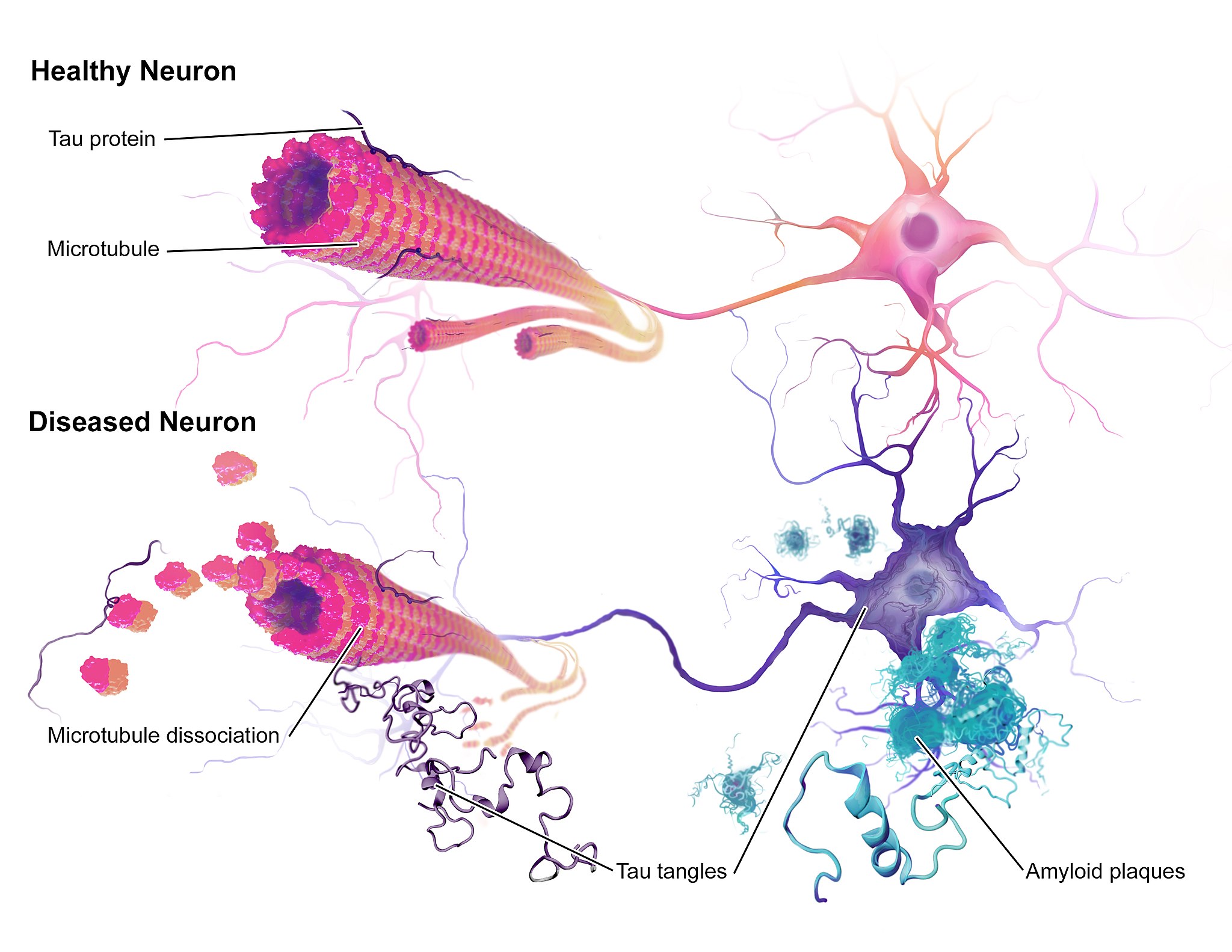
The illustration above (Figure 8.9.1) shows some of the molecular and cellular changes that occur in Alzheimer’s disease (AD). Rosa was diagnosed with AD at the beginning of this chapter after experiencing memory problems and other changes in her cognitive functioning, mood, and personality. These abnormal changes in the brain include the development of amyloid plaques between brain cells and neurofibrillary tangles inside of neurons. These hallmark characteristics of AD are associated with the loss of synapses between neurons, and ultimately the death of neurons.
After reading this chapter, you should have a good appreciation for the importance of keeping neurons alive and communicating with each other at synapses. The nervous system coordinates all of the body’s voluntary and involuntary activities. It interprets information from the outside world through sensory systems, and makes appropriate responses through the motor system, through communication between the PNS and CNS. The brain directs the rest of the nervous system and controls everything from basic vital functions (such as heart rate and breathing) to high-level functions (such as problem solving and abstract thought). The nervous system can perform these important functions by generating action potentials in neurons in response to stimulation and sending messages between cells at synapses, typically using chemical neurotransmitter molecules. When neurons are not functioning properly, lose their synapses, or die, they cannot carry out the signaling essential for the proper functioning of the nervous system.
AD is a progressive neurodegenerative disease, meaning that the damage to the brain becomes more extensive as time goes on. The picture in Figure 8.9.2 illustrates how the damage progresses from before AD is diagnosed (preclinical AD), to mild and moderate AD, to severe AD.
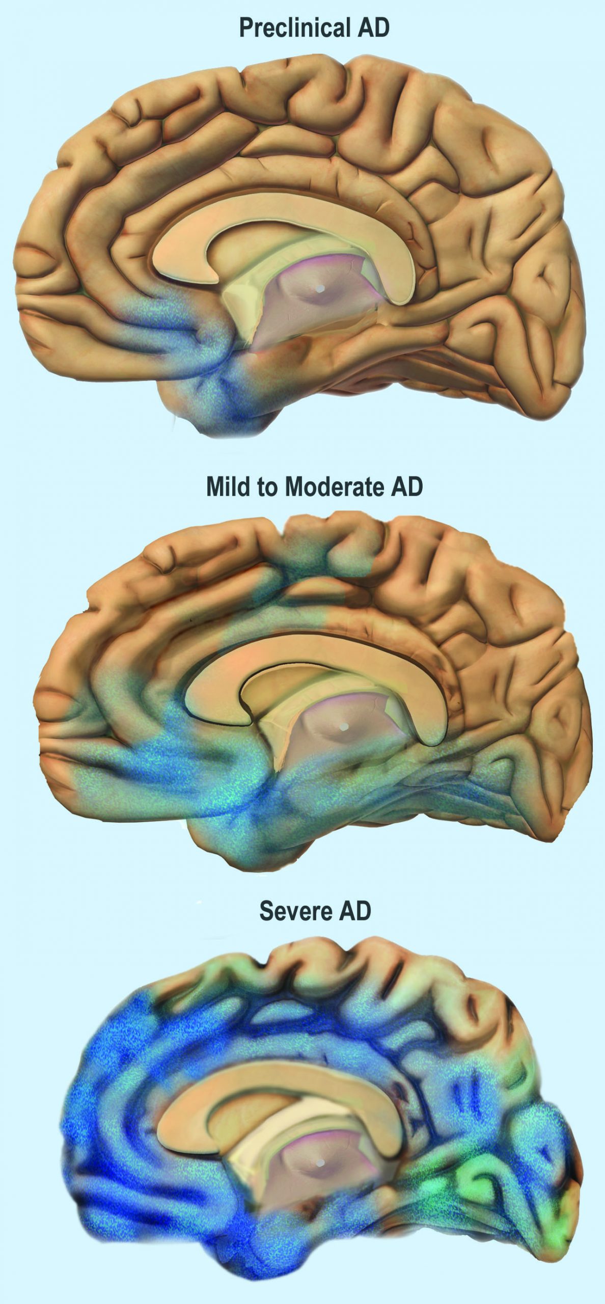
You can see that the damage starts in a relatively small location toward the bottom of the brain. One of the earliest brain areas to be affected by AD is the hippocampus. As you have learned, the hippocampus is important for learning and memory, which explains why many of Rosa’s symptoms of mild AD involve deficits in memory, such as trouble remembering where she placed objects, recent conversations, and appointments.
As AD progresses, more of the brain is affected, including areas involved in emotional regulation, social behavior, planning, language, spatial navigation, and higher-level thought. Rosa is beginning to show signs of problems in these areas, including irritability, lashing out at family members, getting lost in her neighborhood, problems finding the right words, putting objects in unusual locations, and difficulty in managing her finances. You can see that as AD progresses, damage spreads further across the cerebrum, which you now know controls conscious functions like reasoning, language, and interpretation of sensory stimuli. You can also see how the frontal lobe — which controls executive functions such as planning, self-control, and abstract thought — becomes increasingly damaged.
Increasing damage to the brain causes corresponding deficits in functioning. In moderate AD, patients have increased memory, language, and cognitive deficits, compared to mild AD. They may not recognize their own family members, and may wander and get lost, engage in inappropriate behaviors, become easily agitated, and have trouble carrying out daily activities such as dressing. In severe AD, much of the brain is affected. Patients usually cannot recognize family members or communicate, and they are often fully dependent on others for their care. They begin to lose the ability to control their basic functions, such as bladder control, bowel control, and proper swallowing. Eventually, AD causes death, usually as a result of this loss of basic functions.
For now, Rosa only has mild AD and is still able to function relatively well with care from her family. The medication her doctor gave her has helped improve some of her symptoms. It is a cholinesterase inhibitor, which blocks an enzyme that normally degrades the neurotransmitter acetylcholine. With more of the neurotransmitter available, more of it can bind to neurotransmitter receptors on postsynaptic cells. Therefore, this drug acts as an agonist for acetylcholine, which enhances communication between neurons in Rosa’s brain. This increase in neuronal communication can help restore some of the functions lost in early Alzheimer’s disease and may slow the progression of symptoms.
But medication such as this is only a short-term measure, and does not halt the progression of the underlying disease. Ideally, the damaged or dead neurons would be replaced by new, functioning neurons. Why does this not happen automatically in the body? As you have learned, neurogenesis is very limited in adult humans, so once neurons in the brain die, they are not normally replaced to any significant extent. Scientists, however, are studying the ways in which neurogenesis might be increased in cases of disease or injury to the brain. They are also investigating the possibility of using stem cell transplants to replace damaged or dead neurons with new neurons. But this research is in very early stages and is not currently a treatment for AD.
One promising area of research is in the development of methods to allow earlier detection and treatment of AD, given that the changes in the brain may actually start ten to 20 years before diagnosis of AD. A radiolabeled chemical called Pittsburgh Compound B (PiB) binds to amyloid plaques in the brain, and in the future, it may be used in conjunction with brain imaging techniques to detect early signs of AD. Scientists are also looking for biomarkers in bodily fluids (such as blood and cerebrospinal fluid) that might indicate the presence of AD before symptoms appear. Finally, researchers are also investigating possible early and subtle symptoms (such as changes in how people move or a loss of smell) to see whether they can be used to identify people who will go on to develop AD. This research is in the early stages, but the hope is that patients can be identified earlier, allowing for earlier and more effective treatment, as well as more planning time for families.
Scientists are also still trying to fully understand the causes of AD, which affects more than five million Americans. Some genetic mutations have been identified as contributors, but environmental factors also appear to be important. With more research into the causes and mechanisms of AD, hopefully a cure can be found, and people like Rosa can live a longer and better life.
Chapter 8 Summary
In this chapter, you learned about the human nervous system. Specifically, you learned that:
- The nervous system is the organ system that coordinates all of the body’s voluntary and involuntary actions by transmitting signals to and from different parts of the body. It has two major divisions: the central nervous system (CNS) and the peripheral nervous system (PNS).
- The CNS includes the brain and spinal cord.
- The PNS consists mainly of nerves that connect the CNS with the rest of the body. It has two major divisions: the somatic nervous system and the autonomic nervous system. These divisions control different types of functions, and often interact with the CNS to carry out these functions. The somatic system controls activities that are under voluntary control. The autonomic system controls activities that are involuntary.
- The autonomic nervous system is further divided into the sympathetic division (which controls the fight-or-flight response), the parasympathetic division (which controls most routine involuntary responses), and the enteric division (which provides local control for digestive processes).
- Signals sent by the nervous system are electrical signals called nerve impulses. They are transmitted by special, electrically excitable cells called neurons, which are one of two major types of cells in the nervous system.
- Neuroglia are the other major type of nervous system cells. There are many types of glial cells, and they have many specific functions. In general, neuroglia function to support, protect, and nourish neurons.
- The main parts of a neuron include the cell body, dendrites, and axon. The cell body contains the nucleus. Dendrites receive nerve impulses from other cells, and the axon transmits nerve impulses to other cells at axon terminals. A synapse is a complex membrane junction at the end of an axon terminal that transmits signals to another cell.
- Axons are often wrapped in an electrically-insulating myelin sheath, which is produced by oligodendrocytes or schwann cells, both of which are types of neuroglia. Electrical impulses called action potentials occur at gaps in the myelin sheath, called nodes of Ranvier, which speeds the conduction of nerve impulses down the axon.
- Neurogenesis, or the formation of new neurons by cell division, may occur in a mature human brain — but only to a limited extent.
- The nervous tissue in the brain and spinal cord consists of gray matter — which contains mainly unmyelinated cell bodies and dendrites of neurons — and white matter, which contains mainly myelinated axons of neurons. Nerves of the peripheral nervous system consist of long bundles of myelinated axons that extend throughout the body.
- There are hundreds of types of neurons in the human nervous system, but many can be classified on the basis of the direction in which they carry nerve impulses. Sensory neurons carry nerve impulses away from the body and toward the central nervous system, motor neurons carry them away from the central nervous system and toward the body, and interneurons often carry them between sensory and motor neurons.
- A nerve impulse is an electrical phenomenon that occurs because of a difference in electrical charge across the plasma membrane of a neuron.
- The sodium-potassium pump maintains an electrical gradient across the plasma membrane of a neuron when it is not actively transmitting a nerve impulse. This gradient is called the resting potential of the neuron.
- An action potential is a sudden reversal of the electrical gradient across the plasma membrane of a resting neuron. It begins when the neuron receives a chemical signal from another cell or some other type of stimulus. The action potential travels rapidly down the neuron’s axon as an electric current.
- A nerve impulse is transmitted to another cell at either an electrical or a chemical synapse. At a chemical synapse, neurotransmitter chemicals are released from the presynaptic cell into the synaptic cleft between cells. The chemicals travel across the cleft to the postsynaptic cell and bind to receptors embedded in its membrane.
- There are many different types of neurotransmitters. Their effects on the postsynaptic cell generally depend on the type of receptor they bind to. The effects may be excitatory, inhibitory, or modulatory in more complex ways. Both physical and mental disorders may occur if there are problems with neurotransmitters or their receptors.
- The CNS includes the brain and spinal cord. It is physically protected by bones, meninges, and cerebrospinal fluid. It is chemically protected by the blood-brain barrier.
- The brain is the control center of the nervous system and of the entire organism. The brain uses a relatively large proportion of the body’s energy, primarily in the form of glucose.
-
- The brain is divided into three major parts, each with different functions: the forebrain, the midbrain and the hindbrain.
- The forebrain includes the cerebrum, the thalamus, the hypothalamus, the hippocampus and the amygdala. The cerebrum is further divided into left and right hemispheres. Each hemisphere has four lobes: frontal, parietal, temporal, and occipital. Each lobe is associated with specific senses or other functions. The cerebrum has a thin outer layer called the cerebral cortex. Its many folds give it a large surface area. This is where most information processing takes place.
- The thalamus, hypothalamus, hippocampus and amygdala are all part of the limbic system which helps regulate memories, coordination and attention
- The brain is divided into three major parts, each with different functions: the forebrain, the midbrain and the hindbrain.
- The spinal cord is a tubular bundle of nervous tissues that extends from the head down the middle of the back to the pelvis. It functions mainly to connect the brain with the PNS. It also controls certain rapid responses called reflexes without input from the brain.
- A spinal cord injury may lead to paralysis (loss of sensation and movement) of the body below the level of the injury, because nerve impulses can no longer travel up and down the spinal cord beyond that point.
- The PNS consists of all the nervous tissue that lies outside of the CNS. Its main function is to connect the CNS to the rest of the organism.
- The tissues that make up the PNS are nerves and ganglia. Nerves are bundles of axons and ganglia are groups of cell bodies. Nerves are classified as sensory, motor, or a mix of the two.
- The PNS is not as well protected physically or chemically as the CNS, so it is more prone to injury and disease. PNS problems include injury from diabetes, shingles, and heavy metal poisoning. Two disorders of the PNS are Guillain-Barre syndrome and Charcot-Marie-Tooth disease.
- The human body has two major types of senses: special senses and general senses. Special senses have specialized sense organs and include vision (eyes), hearing (ears), balance (ears), taste (tongue), and smell (nasal passages). General senses are all associated with touch and lack special sense organs. Touch receptors are found throughout the body but particularly in the skin.
- All senses depend on sensory receptor cells to detect sensory stimuli and transform them into nerve impulses. Types of sensory receptors include mechanoreceptors (mechanical forces), thermoreceptors (temperature), nociceptors (pain), photoreceptors (light), and chemoreceptors (chemicals).
- Touch includes the ability to sense pressure, vibration, temperature, pain, and other tactile stimuli. The skin includes several different types of touch receptor cells.
- Vision is the ability to sense light and see. The eye is the special sensory organ that collects and focuses light, forms images, and changes them to nerve impulses. Optic nerves send information from the eyes to the brain, which processes the visual information and “tells” us what we are seeing.
- Common vision problems include myopia (nearsightedness), hyperopia (farsightedness), and presbyopia (age-related decline in close vision).
- Hearing is the ability to sense sound waves, and the ear is the organ that senses sound. It changes sound waves to vibrations that trigger nerve impulses, which travel to the brain through the auditory nerve. The brain processes the information and “tells” us what we are hearing.
- The ear is also the organ responsible for the sense of balance, which is the ability to sense and maintain an appropriate body position. The ears send impulses on head position to the brain, which sends messages to skeletal muscle via the peripheral nervous system. The muscles respond by contracting to maintain balance.
- Taste and smell are both abilities to sense chemicals. Taste receptors in taste buds on the tongue sense chemicals in food, and olfactory receptors in the nasal passages sense chemicals in the air. The sense of smell contributes significantly to the sense of taste.
- Psychoactive drugs are substances that change the function of the brain and result in alterations of mood, thinking, perception, and behavior. They include prescription medications (such as opioid painkillers), legal substances (such as nicotine and alcohol), and illegal drugs (such as LSD and heroin).
- Psychoactive drugs are divided into different classes according to their pharmacological effects. They include stimulants, depressants, anxiolytics, euphoriants, hallucinogens, and empathogens. Many psychoactive drugs have multiple effects, so they may be placed in more than one class.
- Psychoactive drugs generally produce their effects by affecting brain chemistry. Generally, they act either as agonists, which enhance the activity of particular neurotransmitters, or as antagonists, which decrease the activity of particular neurotransmitters.
- Psychoactive drugs are used for medical, ritual, and recreational purposes.
- Misuse of psychoactive drugs may lead to addiction, which is the compulsive use of a drug, despite its negative consequences. Sustained use of an addictive drug may produce physical or psychological dependence on the drug. Rehabilitation typically involves psychotherapy, and sometimes the temporary use of other psychoactive drugs.
In addition to the nervous system, there is another system of the body that is important for coordinating and regulating many different functions – the endocrine system. You will learn about the endocrine system in the next chapter.
Chapter 8 Review
- Imagine that you decide to make a movement. To carry out this decision, a neuron in the cerebral cortex of your brain (neuron A) fires a nerve impulse that is sent to a neuron in your spinal cord (neuron B). Neuron B then sends the signal to a muscle cell, causing it to contract, resulting in movement. Answer the following questions about this pathway.
- Which part of the brain is neuron A located in — the cerebellum, cerebrum, or brain stem? Explain how you know.
- The cell body of neuron A is located in a lobe of the brain that is involved in abstract thought, problem solving, and planning. Which lobe is this?
- Part of neuron A travels all the way down to the spinal cord to meet neuron B. Which part of neuron A travels to the spinal cord?
- Neuron A forms a chemical synapse with neuron B in the spinal cord. How is the signal from neuron A transmitted to neuron B?
- Is neuron A in the central nervous system (CNS) or peripheral nervous system (PNS)?
- The axon of neuron B travels in a nerve to a skeletal muscle cell. Is the nerve part of the CNS or PNS? Is this an afferent nerve or an efferent nerve?
- What part of the PNS is involved in this pathway — the autonomic nervous system or the somatic nervous system? Explain your answer.
- What are the differences between a neurotransmitter receptor and a sensory receptor?
-
- If a person has a stroke and then has trouble using language correctly, which hemisphere of their brain was most likely damaged? Explain your answer.
- Electrical gradients are responsible for the resting potential and action potential in neurons. Answer the following questions about the electrical characteristics of neurons.
- Define an electrical gradient, in the context of a cell.
- What is responsible for maintaining the electrical gradient that results in the resting potential?
- Compare and contrast the resting potential and the action potential.
- Where along a myelinated axon does the action potential occur? Why does it happen here?
- What does it mean that the action potential is “all-or-none?”
- Compare and contrast Schwann cells and oligodendrocytes.
- For the senses of smell and hearing, name their respective sensory receptor cells, what type of receptor cells they are, and what stimuli they detect.
- Nicotine is a psychoactive drug that binds to and activates a receptor for the neurotransmitter acetylcholine. Is nicotine an agonist or an antagonist for acetylcholine? Explain your answer.
Attributions
Figure 8.9.1
Alzheimers_Disease by BruceBlaus on Wikimedia Commons is used under a CC BY-SA 4.0 (https://creativecommons.org/licenses/by-sa/4.0/deed.en) license.
Figure 8.9.2
Alzheimer’s Disease stagess by NIH Image Gallery on Flickr is in the public domain (https://en.wikipedia.org/wiki/Public_domain).
The highly complex body system of an animal that coordinates its actions and sensory information by transmitting signals to and from different parts of its body. The nervous system detects environmental changes that impact the body, then works in tandem with the endocrine system to respond to such events.
Actions which take place according to the one's desire or are under control.
Actions which are not under one's conscious control.
One of two main divisions of the nervous system that includes the brain and spinal cord.
One of two major divisions of the nervous system that consists of all the nervous tissue that lies outside the central nervous system.
A division of the peripheral nervous system that controls voluntary activities.
division of the peripheral nervous system that controls involuntary activities
The division of the autonomic nervous system that controls the fight-or-flight response.
The division of the autonomic nervous system that returns the body to normal after the fight-or-flight response and maintains homeostasis at other times.
A division of the autonomic nervous system that controls digestive functions.
A signal transmitted along a nerve fiber.
A functional unit of the nervous system that transmits nerve impulses; also called a nerve cell.
A class of nervous system cell that provides support for neurons and helps them transmit nerve impulses.
The central part of a neuron that contains the nucleus and other cell organelles.
An extension of the cell body of a neuron that receives nerve impulses from other neurons. A neuron will have several dendrites extending from the cell body.
A long extension of the cell body of a neuron that transmits nerve impulses to other cells.
The lipid layer around the axon of a neuron that allows nerve impulses to travel more rapidly down the axon.
Reversal of electrical charge across the membrane of a resting neuron that travels down the axon of the neuron as a nerve impulse.
One of the regularly spaced gaps in the myelin sheath along an axon that allows the action potential (electrical signal) to travel very rapidly.
The formation of new neurons by cell division.
The central nervous system organ inside the skull that is the control center of the nervous system.
A thin, tubular bundle of central nervous system tissue that extends from the brainstem down the back to the pelvis and connects the brain with the peripheral nervous system.
Type of neuron that carries nerve impulses from sensory receptors in tissues and organs to the central nervous system; also called afferent neuron.
A type of neuron that carries nerve impulses from the central nervous system to muscles and glands; also called efferent neuron.
A type of neuron that carries nerve impulses between other neurons, often between sensory and motor neurons.
A solute pump that pumps potassium into cells while pumping sodium out of cells, both against their concentration gradients. This pumping is active and occurs at the ratio of 2 potassium for every 3 calcium.
The place where the axon terminal of a neuron transmits a chemical or electrical signal to another cell.
A type of chemical that transmits signals from the axon of a neuron to another cell across a synapse.
The cell that sends the nerve impulse.
The cell that receives the nerve impulse.
A rigid organ that constitutes part of the vertebrate skeleton in animals.
A three-layered membrane that encloses and protects the brain and spinal cord and contains cerebrospinal fluid.
Clear fluid produced by the brain that forms a thin layer within the meninges and provides protection and cushioning for the brain and spinal cord.
Glucose (also called dextrose) is a simple sugar with the molecular formula C6H12O6. Glucose is the most abundant monosaccharide, a subcategory of carbohydrates. Glucose is mainly made by plants and most algae during photosynthesis from water and carbon dioxide, using energy from sunlight.
The largest part of the brain that controls conscious functions such as reasoning and sight.
The inner part of the brain that is a major hub for nerve impulses traveling back and forth between the cerebrum and spinal cord.
A part of the brain that secretes hormones and connects the brain with the endocrine system.
A complex brain structure embedded deep into temporal lobe. It has a major role in learning and memory.
A roughly almond-shaped mass of gray matter inside each cerebral hemisphere, involved with the experiencing of emotions. Responsible for the perception of emotions such as anger, fear, and sadness, as well as the controlling of aggression. The amygdala helps to store memories of events and emotions so that an individual may be able to recognize similar events in the future.
A structure in the nervous system that consists of cable-like bundles of axons and makes up the majority of the peripheral nervous system.
A structure containing neuronal cell bodies in the peripheral nervous system.
A type of sensory receptor that responds to mechanical forces.
A type of sensory receptor that senses temperature.
A type of sensory receptor that responds to pain.
A type of sensory receptor that responds to light.
A type of sensory receptor that responds to presence of chemicals.
The ability to sense pressure, vibration, temperature, pain, and other tactile stimuli.
The ability to sense light and see; also called sight.
A vision problem in which distant objects are out of focus but close vision is unaffected; also called nearsightedness.
A vision problem in which close objects are out of focus but distant vision is unaffected; also called farsightedness.
Farsightedness caused by loss of elasticity of the lens of the eye, occurring typically in middle and old age.
The ability to sense sound waves.
The ability to sense and maintain an appropriate body position.
The perception produced or stimulated when a substance in the mouth reacts chemically with taste receptor cells located on taste buds in the oral cavity, mostly on the tongue.
A chemoreception (ability to detect the presence of certain chemicals) that, through the sensory olfactory system, forms the perception of smell.
A drug that affects the central nervous system, generally by influencing neurotransmitters in the brain.
A type of psychoactive drug that stimulates the brain and increases alertness and wakefulness.
A type of psychoactive drug that calms the brain, reduces anxious feelings, and induces sleepiness.
Type of psychoactive drug that has a tranquilizing effect and inhibits anxiety.
A type of psychoactive drug that brings about a state of euphoria.
A type of psychoactive drug that causes hallucinations and other perceptual anomalies, as well as subjective changes in thoughts, emotions, and consciousness.
A type of psychoactive drug that produces feelings of empathy with other people.
A drug that increases the activity or effect of a neurotransmitter.
A drug that decreases the activity of a particular neurotransmitter.
The compulsive use of a drug, despite negative consequences that such use may entail.
A state of reliance upon a drug such that when the drug is withdrawn, several physiologic reactions occur.
