
28 4.2 Discovery of Cells and Cell Theory
Created by: CK-12/Adapted by Christine Miller
Figure 4.2.1 Human cells viewed with a very powerful tool called a scanning electron microscope.
Amazing Cells
What are these incredible objects? Would it surprise you to learn that they are all human cells? Cells are actually too small to see with the unaided eye. It is visible here in such detail because it is being viewed with a very powerful tool called a scanning electron microscope. Cells may be small in size, but they are extremely important to life. Like all other living things, you are made of cells. Cells are the basis of life, and without cells, life as we know it would not exist. You will learn more about these amazing building blocks of life in this section.
What Are Cells?
If you look at living matter with a microscope — even a simple light microscope — you will see that it consists of cells. Cells are the basic units of the structure and function of living things. They are the smallest units that can carry out the processes of life. All organisms are made up of one or more cells, and all cells have many of the same structures and carry out the same basic life processes. Knowing the structure of cells and the processes they carry out is necessary to an understanding of life itself.
Discovery of Cells
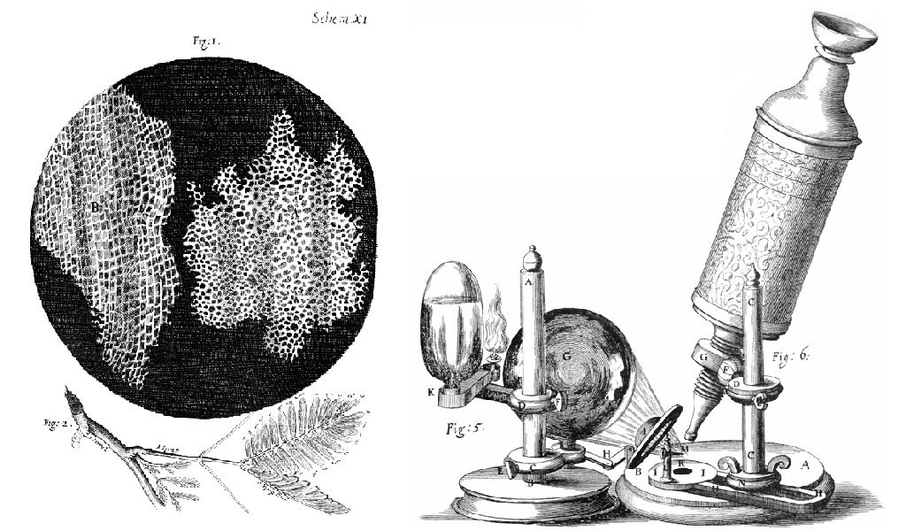
The first time the word cell was used to refer to these tiny units of life was in 1665 by a British scientist named Robert Hooke. Hooke was one of the earliest scientists to study living things under a microscope. The microscopes of his day were not very strong, but Hooke was still able to make an important discovery. When he looked at a thin slice of cork under his microscope, he was surprised to see what looked like a honeycomb. Hooke made the drawing in the figure to the right to show what he saw. As you can see, the cork was made up of many tiny units. Hooke called these units cells because they resembled cells in a monastery.
Soon after Robert Hooke discovered cells in cork, Anton van Leeuwenhoek in Holland made other important discoveries using a microscope. Leeuwenhoek made his own microscope lenses, and he was so good at it that his microscope was more powerful than other microscopes of his day. In fact, Leeuwenhoek’s microscope was almost as strong as modern light microscopes. Using his microscope, Leeuwenhoek was the first person to observe human cells and bacteria.
Cell Theory
By the early 1800s, scientists had observed cells of many different organisms. These observations led two German scientists named Theodor Schwann and Matthias Jakob Schleiden to propose cells as the basic building blocks of all living things. Around 1850, a German doctor named Rudolf Virchow was studying cells under a microscope, when he happened to see them dividing and forming new cells. He realized that living cells produce new cells through division. Based on this realization, Virchow proposed that living cells arise only from other living cells.
The ideas of all three scientists — Schwann, Schleiden, and Virchow — led to cell theory, which is one of the fundamental theories unifying all of biology.
Cell theory states that:
- All organisms are made of one or more cells.
- All the life functions of organisms occur within cells.
- All cells come from existing cells.
Seeing Inside Cells
Starting with Robert Hooke in the 1600s, the microscope opened up an amazing new world — a world of life at the level of the cell. As microscopes continued to improve, more discoveries were made about the cells of living things, but by the late 1800s, light microscopes had reached their limit. Objects much smaller than cells (including the structures inside cells) were too small to be seen with even the strongest light microscope.
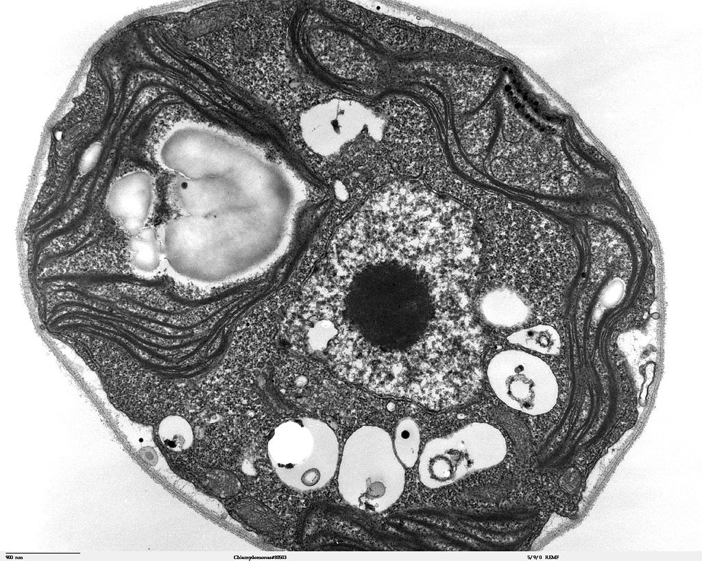
Then, in the 1950s, a new type of microscope was invented. Called the electron microscope, it used a beam of electrons instead of light to observe extremely small objects. With an electron microscope, scientists could finally see the tiny structures inside cells. They could even see individual molecules and atoms. The electron microscope had a huge impact on biology. It allowed scientists to study organisms at the level of their molecules, and it led to the emergence of the molecular biology field. With the electron microscope, many more cell discoveries were made.
Structures Shared By All Cells
Although cells are diverse, all cells have certain parts in common. These parts include a plasma membrane, cytoplasm, ribosomes, and DNA.
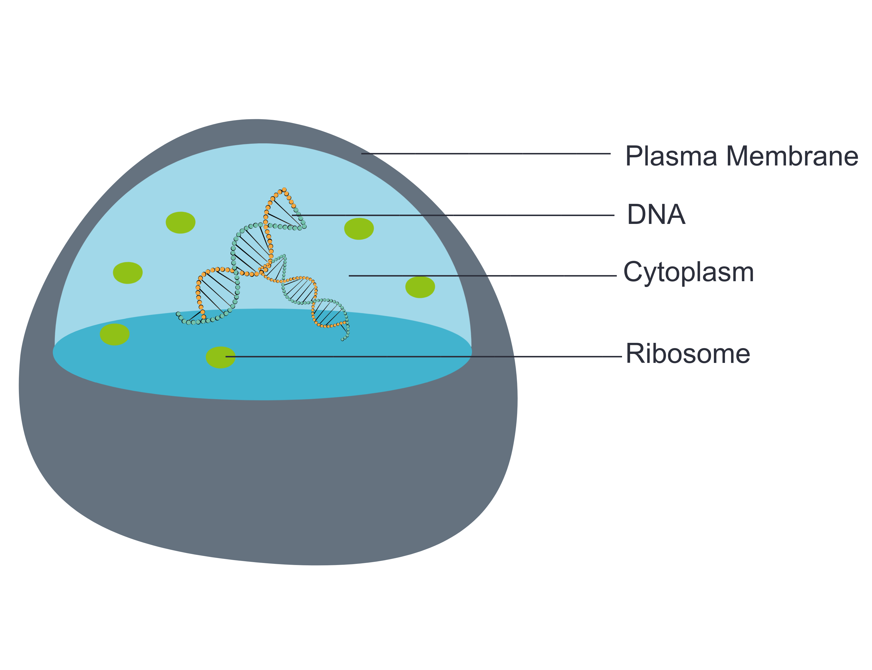
- The plasma membrane (a type of cell membrane) is a thin coat of lipids that surrounds a cell. It forms the physical boundary between the cell and its environment. You can think of it as the “skin” of the cell.
- Cytoplasm refers to all of the cellular material inside of the plasma membrane. Cytoplasm is made up of a watery substance called cytosol, and it contains other cell structures, such as ribosomes.
- Ribosomes are the structures in the cytoplasm in which proteins are made.
- DNA is a nucleic acid found in cells. It contains the genetic instructions that cells need to make proteins.
These four parts are common to all cells, from organisms as different as bacteria and human beings. How did all known organisms come to have such similar cells? The similarities show that all life on Earth has a common evolutionary history.
4.2 Summary
- Cells are the basic units of structure and function in living things. They are the smallest units that can carry out the processes of life.
- In the 1600s, Hooke was the first to observe cells from an organism (cork). Soon after, microscopist van Leeuwenhoek observed many other living cells.
- In the early 1800s, Schwann and Schleiden theorized that cells are the basic building blocks of all living things. Around 1850, Virchow observed cells dividing. To previous learnings, he added that living cells arise only from other living cells. These ideas led to cell theory, which states that all organisms are made of cells, that all life functions occur in cells, and that all cells come from other cells.
- It wasn’t until the 1950s that scientists could see what was inside the cell. The invention of the electron microscope allowed them to see organelles and other structures smaller than cells.
- There is variation in cells, but all cells have a plasma membrane, cytoplasm, ribosomes, and DNA. These similarities show that all life on Earth has a common ancestor in the distant past.
4.2 Review Questions
- Describe cells.
- Explain how cells were discovered.
- Outline the development of cell theory.
-
- Identify the structures shared by all cells.
- Proteins are made on _____________ .
-
- Robert Hooke sketched what looked like honeycombs — or repeated circular or square units — when he observed plant cells under a microscope.
- What is each unit?
- Of the shared parts of all cells, what makes up the outer surface of each unit?
- Of the shared parts of all cells, what makes up the inside of each unit?
4.2 Explore More
Introduction to Cells: The Grand Cell Tour, by The Amoeba Sisters, 2016.
Attributions
Figure 4.2.1
- A white blood cell (WBC) known as a neutrophil by National Institute of Allergy and Infectious Diseases (NIAID) on the CDC/ Public Health Image Library (PHIL) Photo ID# 18129. is in the public domain (https://en.wikipedia.org/wiki/Public_domain).
- Healthy Human T Cell by NIAID on Flickr. is used under a CC BY 2.0 (https://creativecommons.org/licenses/by/2.0/) license.
- Human natural killer cell by NIAID on Flickr. is used under a CC BY 2.0 (https://creativecommons.org/licenses/by/2.0/) license.
- Human blood with red blood cells, T cells (orange) and platelets (green) by ZEISS Microscopy on Flickr. is used under a CC BY-NC-ND 2.0 (https://creativecommons.org/licenses/by-nc-nd/2.0/) license.
- Developing nerve cells by ZEISS Microscopy on Flickr. is used under a CC BY-NC-ND 2.0 (https://creativecommons.org/licenses/by-nc-nd/2.0/) license.
Figure 4.2.2
Hooke-microscope-cork by Robert Hooke (1635-1702) [uploaded by Alejandro Porto] on Wikimedia Commons is released into the public domain (https://en.wikipedia.org/wiki/Public_domain).
Figure 4.2.3
Electron Microscope image of a cell by Dartmouth Electron Microscope Facility, Dartmouth College on Wikimedia Commons is released into the public domain (https://en.wikipedia.org/wiki/Public_domain).
Figure 4.2.4
Basic-Components-of-a-cell by Christine Miller is used under a CC0 1.0 (https://creativecommons.org/publicdomain/zero/1.0/) license.
References
Amoeba Sisters. (2016, November 1). Introduction to cells: The grand cell tour. YouTube. https://www.youtube.com/watch?v=8IlzKri08kk&feature=youtu.be
National Institute of Allergy and Infectious Diseases (NIAID). (2011). A white blood cell (WBC) known as a neutrophil, as it was in the process of ingesting a number of spheroid shaped, methicillin-resistant, Staphylococcus aureus (MRSA) bacteria [digital image]. CDC/ Public Health Image Library (PHIL) Photo ID# 18129. https://phil.cdc.gov/Details.aspx?pid=18129.
Wikipedia contributors. (2020, June 24). Antonie van Leeuwenhoek. In Wikipedia. https://en.wikipedia.org/w/index.php?title=Antonie_van_Leeuwenhoek&oldid=964339564
Wikipedia contributors. (2020, May 25). Matthias Jakob Schleiden. In Wikipedia. https://en.wikipedia.org/w/index.php?title=Matthias_Jakob_Schleiden&oldid=958819219
Wikipedia contributors. (2020, June 4). Rudolf Virchow. In Wikipedia,. https://en.wikipedia.org/w/index.php?title=Rudolf_Virchow&oldid=960708716
Wikipedia contributors. (2020, May 16). Theodor Schwann. In Wikipedia. https://en.wikipedia.org/w/index.php?title=Theodor_Schwann&oldid=956919239
The smallest unit of life, consisting of at least a membrane, cytoplasm, and genetic material.
A historic scientific theory consisting of 3 statements: all living organisms of made of one or more cells, the cell is the basic unit of all living things, and all cells arise from pre-existing cells.
A semi-permeable lipid bilayer that separates the interior of all cells from their surroundings.
The jellylike material that makes up much of a cell inside the cell membrane, and, in eukaryotic cells, surrounds the nucleus. The organelles of eukaryotic cells, such as mitochondria, the endoplasmic reticulum, and (in green plants) chloroplasts, are contained in the cytoplasm.
A large complex of RNA and protein which acts as the site of RNA translation, building proteins from amino acids using messenger RNA as a template.
Deoxyribonucleic acid - the molecule carrying genetic instructions for the development, functioning, growth and reproduction of all known organisms and many viruses.
